Deyun Zhang
ECG-R1: Protocol-Guided and Modality-Agnostic MLLM for Reliable ECG Interpretation
Feb 04, 2026Abstract:Electrocardiography (ECG) serves as an indispensable diagnostic tool in clinical practice, yet existing multimodal large language models (MLLMs) remain unreliable for ECG interpretation, often producing plausible but clinically incorrect analyses. To address this, we propose ECG-R1, the first reasoning MLLM designed for reliable ECG interpretation via three innovations. First, we construct the interpretation corpus using \textit{Protocol-Guided Instruction Data Generation}, grounding interpretation in measurable ECG features and monograph-defined quantitative thresholds and diagnostic logic. Second, we present a modality-decoupled architecture with \textit{Interleaved Modality Dropout} to improve robustness and cross-modal consistency when either the ECG signal or ECG image is missing. Third, we present \textit{Reinforcement Learning with ECG Diagnostic Evidence Rewards} to strengthen evidence-grounded ECG interpretation. Additionally, we systematically evaluate the ECG interpretation capabilities of proprietary, open-source, and medical MLLMs, and provide the first quantitative evidence that severe hallucinations are widespread, suggesting that the public should not directly trust these outputs without independent verification. Code and data are publicly available at \href{https://github.com/PKUDigitalHealth/ECG-R1}{here}, and an online platform can be accessed at \href{http://ai.heartvoice.com.cn/ECG-R1/}{here}.
Aortic Valve Disease Detection from PPG via Physiology-Informed Self-Supervised Learning
Feb 04, 2026Abstract:Traditional diagnosis of aortic valve disease relies on echocardiography, but its cost and required expertise limit its use in large-scale early screening. Photoplethysmography (PPG) has emerged as a promising screening modality due to its widespread availability in wearable devices and its ability to reflect underlying hemodynamic dynamics. However, the extreme scarcity of gold-standard labeled PPG data severely constrains the effectiveness of data-driven approaches. To address this challenge, we propose and validate a new paradigm, Physiology-Guided Self-Supervised Learning (PG-SSL), aimed at unlocking the value of large-scale unlabeled PPG data for efficient screening of Aortic Stenosis (AS) and Aortic Regurgitation (AR). Using over 170,000 unlabeled PPG samples from the UK Biobank, we formalize clinical knowledge into a set of PPG morphological phenotypes and construct a pulse pattern recognition proxy task for self-supervised pre-training. A dual-branch, gated-fusion architecture is then employed for efficient fine-tuning on a small labeled subset. The proposed PG-SSL framework achieves AUCs of 0.765 and 0.776 for AS and AR screening, respectively, significantly outperforming supervised baselines trained on limited labeled data. Multivariable analysis further validates the model output as an independent digital biomarker with sustained prognostic value after adjustment for standard clinical risk factors. This study demonstrates that PG-SSL provides an effective, domain knowledge-driven solution to label scarcity in medical artificial intelligence and shows strong potential for enabling low-cost, large-scale early screening of aortic valve disease.
ECGomics: An Open Platform for AI-ECG Digital Biomarker Discovery
Jan 19, 2026Abstract:Background: Conventional electrocardiogram (ECG) analysis faces a persistent dichotomy: expert-driven features ensure interpretability but lack sensitivity to latent patterns, while deep learning offers high accuracy but functions as a black box with high data dependency. We introduce ECGomics, a systematic paradigm and open-source platform for the multidimensional deconstruction of cardiac signals into digital biomarker. Methods: Inspired by the taxonomic rigor of genomics, ECGomics deconstructs cardiac activity across four dimensions: Structural, Intensity, Functional, and Comparative. This taxonomy synergizes expert-defined morphological rules with data-driven latent representations, effectively bridging the gap between handcrafted features and deep learning embeddings. Results: We operationalized this framework into a scalable ecosystem consisting of a web-based research platform and a mobile-integrated solution (https://github.com/PKUDigitalHealth/ECGomics). The web platform facilitates high-throughput analysis via precision parameter configuration, high-fidelity data ingestion, and 12-lead visualization, allowing for the systematic extraction of biomarkers across the four ECGomics dimensions. Complementarily, the mobile interface, integrated with portable sensors and a cloud-based engine, enables real-time signal acquisition and near-instantaneous delivery of structured diagnostic reports. This dual-interface architecture successfully transitions ECGomics from theoretical discovery to decentralized, real-world health management, ensuring professional-grade monitoring in diverse clinical and home-based settings. Conclusion: ECGomics harmonizes diagnostic precision, interpretability, and data efficiency. By providing a deployable software ecosystem, this paradigm establishes a robust foundation for digital biomarker discovery and personalized cardiovascular medicine.
AnyECG: Evolved ECG Foundation Model for Holistic Health Profiling
Jan 12, 2026Abstract:Background: Artificial intelligence enabled electrocardiography (AI-ECG) has demonstrated the ability to detect diverse pathologies, but most existing models focus on single disease identification, neglecting comorbidities and future risk prediction. Although ECGFounder expanded cardiac disease coverage, a holistic health profiling model remains needed. Methods: We constructed a large multicenter dataset comprising 13.3 million ECGs from 2.98 million patients. Using transfer learning, ECGFounder was fine-tuned to develop AnyECG, a foundation model for holistic health profiling. Performance was evaluated using external validation cohorts and a 10-year longitudinal cohort for current diagnosis, future risk prediction, and comorbidity identification. Results: AnyECG demonstrated systemic predictive capability across 1172 conditions, achieving an AUROC greater than 0.7 for 306 diseases. The model revealed novel disease associations, robust comorbidity patterns, and future disease risks. Representative examples included high diagnostic performance for hyperparathyroidism (AUROC 0.941), type 2 diabetes (0.803), Crohn disease (0.817), lymphoid leukemia (0.856), and chronic obstructive pulmonary disease (0.773). Conclusion: The AnyECG foundation model provides substantial evidence that AI-ECG can serve as a systemic tool for concurrent disease detection and long-term risk prediction.
Artificial Intelligence-Enabled Spirometry for Early Detection of Right Heart Failure
Nov 17, 2025Abstract:Right heart failure (RHF) is a disease characterized by abnormalities in the structure or function of the right ventricle (RV), which is associated with high morbidity and mortality. Lung disease often causes increased right ventricular load, leading to RHF. Therefore, it is very important to screen out patients with cor pulmonale who develop RHF from people with underlying lung diseases. In this work, we propose a self-supervised representation learning method to early detecting RHF from patients with cor pulmonale, which uses spirogram time series to predict patients with RHF at an early stage. The proposed model is divided into two stages. The first stage is the self-supervised representation learning-based spirogram embedding (SLSE) network training process, where the encoder of the Variational autoencoder (VAE-encoder) learns a robust low-dimensional representation of the spirogram time series from the data-augmented unlabeled data. Second, this low-dimensional representation is fused with demographic information and fed into a CatBoost classifier for the downstream RHF prediction task. Trained and tested on a carefully selected subset of 26,617 individuals from the UK Biobank, our model achieved an AUROC of 0.7501 in detecting RHF, demonstrating strong population-level distinction ability. We further evaluated the model on high-risk clinical subgroups, achieving AUROC values of 0.8194 on a test set of 74 patients with chronic kidney disease (CKD) and 0.8413 on a set of 64 patients with valvular heart disease (VHD). These results highlight the model's potential utility in predicting RHF among clinically elevated-risk populations. In conclusion, this study presents a self-supervised representation learning approach combining spirogram time series and demographic data, demonstrating promising potential for early RHF detection in clinical practice.
AnyECG-Lab: An Exploration Study of Fine-tuning an ECG Foundation Model to Estimate Laboratory Values from Single-Lead ECG Signals
Oct 25, 2025

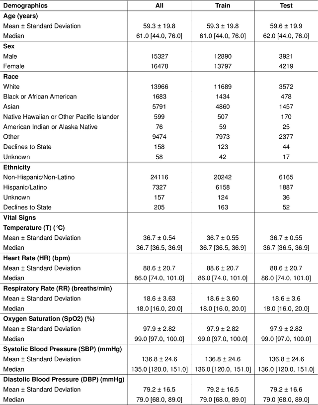
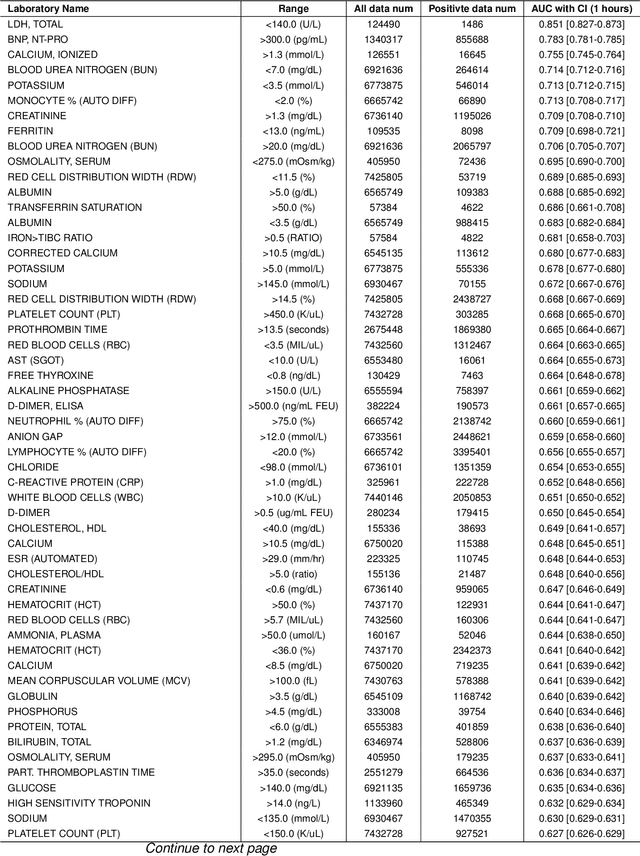
Abstract:Timely access to laboratory values is critical for clinical decision-making, yet current approaches rely on invasive venous sampling and are intrinsically delayed. Electrocardiography (ECG), as a non-invasive and widely available signal, offers a promising modality for rapid laboratory estimation. Recent progress in deep learning has enabled the extraction of latent hematological signatures from ECGs. However, existing models are constrained by low signal-to-noise ratios, substantial inter-individual variability, limited data diversity, and suboptimal generalization, especially when adapted to low-lead wearable devices. In this work, we conduct an exploratory study leveraging transfer learning to fine-tune ECGFounder, a large-scale pre-trained ECG foundation model, on the Multimodal Clinical Monitoring in the Emergency Department (MC-MED) dataset from Stanford. We generated a corpus of more than 20 million standardized ten-second ECG segments to enhance sensitivity to subtle biochemical correlates. On internal validation, the model demonstrated strong predictive performance (area under the curve above 0.65) for thirty-three laboratory indicators, moderate performance (between 0.55 and 0.65) for fifty-nine indicators, and limited performance (below 0.55) for sixteen indicators. This study provides an efficient artificial-intelligence driven solution and establishes the feasibility scope for real-time, non-invasive estimation of laboratory values.
Accuracy of Wearable ECG Parameter Calculation Method for Long QT and First-Degree A-V Block Detection: A Multi-Center Real-World Study with External Validations Compared to Standard ECG Machines and Cardiologist Assessments
Feb 21, 2025Abstract:In recent years, wearable devices have revolutionized cardiac monitoring by enabling continuous, non-invasive ECG recording in real-world settings. Despite these advances, the accuracy of ECG parameter calculations (PR interval, QRS interval, QT interval, etc.) from wearables remains to be rigorously validated against conventional ECG machines and expert clinician assessments. In this large-scale, multicenter study, we evaluated FeatureDB, a novel algorithm for automated computation of ECG parameters from wearable single-lead signals Three diverse datasets were employed: the AHMU-FH dataset (n=88,874), the CSE dataset (n=106), and the HeartVoice-ECG-lite dataset (n=369) with annotations provided by two experienced cardiologists. FeatureDB demonstrates a statistically significant correlation with key parameters (PR interval, QRS duration, QT interval, and QTc) calculated by standard ECG machines and annotated by clinical doctors. Bland-Altman analysis confirms a high level of agreement.Moreover,FeatureDB exhibited robust diagnostic performance in detecting Long QT syndrome (LQT) and atrioventricular block interval abnormalities (AVBI),with excellent area under the ROC curve (LQT: 0.836, AVBI: 0.861),accuracy (LQT: 0.856, AVBI: 0.845),sensitivity (LQT: 0.815, AVBI: 0.877),and specificity (LQT: 0.856, AVBI: 0.845).This further validates its clinical reliability. These results validate the clinical applicability of FeatureDB for wearable ECG analysis and highlight its potential to bridge the gap between traditional diagnostic methods and emerging wearable technologies.Ultimately,this study supports integrating wearable ECG devices into large-scale cardiovascular disease management and early intervention strategies,and it highlights the potential of wearable ECG technologies to deliver accurate,clinically relevant cardiac monitoring while advancing broader applications in cardiovascular care.
DiffuSETS: 12-lead ECG Generation Conditioned on Clinical Text Reports and Patient-Specific Information
Jan 10, 2025Abstract:Heart disease remains a significant threat to human health. As a non-invasive diagnostic tool, the electrocardiogram (ECG) is one of the most widely used methods for cardiac screening. However, the scarcity of high-quality ECG data, driven by privacy concerns and limited medical resources, creates a pressing need for effective ECG signal generation. Existing approaches for generating ECG signals typically rely on small training datasets, lack comprehensive evaluation frameworks, and overlook potential applications beyond data augmentation. To address these challenges, we propose DiffuSETS, a novel framework capable of generating ECG signals with high semantic alignment and fidelity. DiffuSETS accepts various modalities of clinical text reports and patient-specific information as inputs, enabling the creation of clinically meaningful ECG signals. Additionally, to address the lack of standardized evaluation in ECG generation, we introduce a comprehensive benchmarking methodology to assess the effectiveness of generative models in this domain. Our model achieve excellent results in tests, proving its superiority in the task of ECG generation. Furthermore, we showcase its potential to mitigate data scarcity while exploring novel applications in cardiology education and medical knowledge discovery, highlighting the broader impact of our work.
A Review of Deep Learning Methods for Photoplethysmography Data
Jan 23, 2024Abstract:Photoplethysmography (PPG) is a highly promising device due to its advantages in portability, user-friendly operation, and non-invasive capabilities to measure a wide range of physiological information. Recent advancements in deep learning have demonstrated remarkable outcomes by leveraging PPG signals for tasks related to personal health management and other multifaceted applications. In this review, we systematically reviewed papers that applied deep learning models to process PPG data between January 1st of 2017 and July 31st of 2023 from Google Scholar, PubMed and Dimensions. Each paper is analyzed from three key perspectives: tasks, models, and data. We finally extracted 193 papers where different deep learning frameworks were used to process PPG signals. Based on the tasks addressed in these papers, we categorized them into two major groups: medical-related, and non-medical-related. The medical-related tasks were further divided into seven subgroups, including blood pressure analysis, cardiovascular monitoring and diagnosis, sleep health, mental health, respiratory monitoring and analysis, blood glucose analysis, as well as others. The non-medical-related tasks were divided into four subgroups, which encompass signal processing, biometric identification, electrocardiogram reconstruction, and human activity recognition. In conclusion, significant progress has been made in the field of using deep learning methods to process PPG data recently. This allows for a more thorough exploration and utilization of the information contained in PPG signals. However, challenges remain, such as limited quantity and quality of publicly available databases, a lack of effective validation in real-world scenarios, and concerns about the interpretability, scalability, and complexity of deep learning models. Moreover, there are still emerging research areas that require further investigation.
Artificial Intelligence System for Detection and Screening of Cardiac Abnormalities using Electrocardiogram Images
Feb 10, 2023


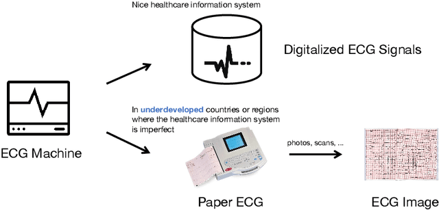
Abstract:The artificial intelligence (AI) system has achieved expert-level performance in electrocardiogram (ECG) signal analysis. However, in underdeveloped countries or regions where the healthcare information system is imperfect, only paper ECGs can be provided. Analysis of real-world ECG images (photos or scans of paper ECGs) remains challenging due to complex environments or interference. In this study, we present an AI system developed to detect and screen cardiac abnormalities (CAs) from real-world ECG images. The system was evaluated on a large dataset of 52,357 patients from multiple regions and populations across the world. On the detection task, the AI system obtained area under the receiver operating curve (AUC) of 0.996 (hold-out test), 0.994 (external test 1), 0.984 (external test 2), and 0.979 (external test 3), respectively. Meanwhile, the detection results of AI system showed a strong correlation with the diagnosis of cardiologists (cardiologist 1 (R=0.794, p<1e-3), cardiologist 2 (R=0.812, p<1e-3)). On the screening task, the AI system achieved AUCs of 0.894 (hold-out test) and 0.850 (external test). The screening performance of the AI system was better than that of the cardiologists (AI system (0.846) vs. cardiologist 1 (0.520) vs. cardiologist 2 (0.480)). Our study demonstrates the feasibility of an accurate, objective, easy-to-use, fast, and low-cost AI system for CA detection and screening. The system has the potential to be used by healthcare professionals, caregivers, and general users to assess CAs based on real-world ECG images.
 Add to Chrome
 Add to Firefox
 Add to Edge