 Add to Chrome
 Add to Firefox
 Add to Edge
MultiCo3D: Multi-Label Voxel Contrast for One-Shot Incremental Segmentation of 3D Neuroimages
Mar 09, 20253D neuroimages provide a comprehensive view of brain structure and function, aiding in precise localization and functional connectivity analysis. Segmentation of white matter (WM) tracts using 3D neuroimages is vital for understanding the brain's structural connectivity in both healthy and diseased states. One-shot Class Incremental Semantic Segmentation (OCIS) refers to effectively segmenting new (novel) classes using only a single sample while retaining knowledge of old (base) classes without forgetting. Voxel-contrastive OCIS methods adjust the feature space to alleviate the feature overlap problem between the base and novel classes. However, since WM tract segmentation is a multi-label segmentation task, existing single-label voxel contrastive-based methods may cause inherent contradictions. To address this, we propose a new multi-label voxel contrast framework called MultiCo3D for one-shot class incremental tract segmentation. Our method utilizes uncertainty distillation to preserve base tract segmentation knowledge while adjusting the feature space with multi-label voxel contrast to alleviate feature overlap when learning novel tracts and dynamically weighting multi losses to balance overall loss. We compare our method against several state-of-the-art (SOTA) approaches. The experimental results show that our method significantly enhances one-shot class incremental tract segmentation accuracy across five different experimental setups on HCP and Preto datasets.
DDCSR: A Novel End-to-End Deep Learning Framework for Cortical Surface Reconstruction from Diffusion MRI
Mar 05, 2025Diffusion MRI (dMRI) plays a crucial role in studying brain white matter connectivity. Cortical surface reconstruction (CSR), including the inner whiter matter (WM) and outer pial surfaces, is one of the key tasks in dMRI analyses such as fiber tractography and multimodal MRI analysis. Existing CSR methods rely on anatomical T1-weighted data and map them into the dMRI space through inter-modality registration. However, due to the low resolution and image distortions of dMRI data, inter-modality registration faces significant challenges. This work proposes a novel end-to-end learning framework, DDCSR, which for the first time enables CSR directly from dMRI data. DDCSR consists of two major components, including: (1) an implicit learning module to predict a voxel-wise intermediate surface representation, and (2) an explicit learning module to predict the 3D mesh surfaces. Compared to several baseline and advanced CSR methods, we show that the proposed DDCSR can largely increase both accuracy and efficiency. Furthermore, we demonstrate a high generalization ability of DDCSR to data from different sources, despite the differences in dMRI acquisitions and populations.
Medical Image Registration Meets Vision Foundation Model: Prototype Learning and Contour Awareness
Feb 17, 2025Medical image registration is a fundamental task in medical image analysis, aiming to establish spatial correspondences between paired images. However, existing unsupervised deformable registration methods rely solely on intensity-based similarity metrics, lacking explicit anatomical knowledge, which limits their accuracy and robustness. Vision foundation models, such as the Segment Anything Model (SAM), can generate high-quality segmentation masks that provide explicit anatomical structure knowledge, addressing the limitations of traditional methods that depend only on intensity similarity. Based on this, we propose a novel SAM-assisted registration framework incorporating prototype learning and contour awareness. The framework includes: (1) Explicit anatomical information injection, where SAM-generated segmentation masks are used as auxiliary inputs throughout training and testing to ensure the consistency of anatomical information; (2) Prototype learning, which leverages segmentation masks to extract prototype features and aligns prototypes to optimize semantic correspondences between images; and (3) Contour-aware loss, a contour-aware loss is designed that leverages the edges of segmentation masks to improve the model's performance in fine-grained deformation fields. Extensive experiments demonstrate that the proposed framework significantly outperforms existing methods across multiple datasets, particularly in challenging scenarios with complex anatomical structures and ambiguous boundaries. Our code is available at https://github.com/HaoXu0507/IPMI25-SAM-Assisted-Registration.
Signal-adapted decomposition of graph signals
Feb 17, 2025Analysis of signals defined on complex topologies modeled by graphs is a topic of increasing interest. Signal decomposition plays a crucial role in the representation and processing of such information, in particular, to process graph signals based on notions of scale (e.g., coarse to fine). The graph spectrum is more irregular than for conventional domains; i.e., it is influenced by graph topology, and, therefore, assumptions about spectral representations of graph signals are not easy to make. Here, we propose a tight frame design that is adapted to the graph Laplacian spectral content of given classes of graph signals. The design is based on using the ensemble energy spectral density, a notion of spectral content of given signal sets that we determine either directly using the graph Fourier transform or indirectly through a polynomial-based approximation scheme. The approximation scheme has the benefit that (i) it does not require eigendecomposition of the Laplacian matrix making the method feasible for large graphs, and (ii) it leads to a smooth estimate of the spectral content. A prototype system of spectral kernels each capturing an equal amount of energy is initially defined and subsequently warped using the signal set's ensemble energy spectral density such that the resulting subbands each capture an equal amount of ensemble energy. This approach accounts at the same time for graph topology and signal features, and it provides a meaningful interpretation of subbands in terms of coarse-to-fine representations. We also show how more simplified designs of signal-adapted decomposition of graph signals can be adopted based on ensemble energy estimates. We show the application of proposed methods on the Minnesota road graph and three different designs of brain graphs derived from neuroimaging data.
EVENet: Evidence-based Ensemble Learning for Uncertainty-aware Brain Parcellation Using Diffusion MRI
Sep 11, 2024


In this study, we developed an Evidence-based Ensemble Neural Network, namely EVENet, for anatomical brain parcellation using diffusion MRI. The key innovation of EVENet is the design of an evidential deep learning framework to quantify predictive uncertainty at each voxel during a single inference. Using EVENet, we obtained accurate parcellation and uncertainty estimates across different datasets from healthy and clinical populations and with different imaging acquisitions. The overall network includes five parallel subnetworks, where each is dedicated to learning the FreeSurfer parcellation for a certain diffusion MRI parameter. An evidence-based ensemble methodology is then proposed to fuse the individual outputs. We perform experimental evaluations on large-scale datasets from multiple imaging sources, including high-quality diffusion MRI data from healthy adults and clinically diffusion MRI data from participants with various brain diseases (schizophrenia, bipolar disorder, attention-deficit/hyperactivity disorder, Parkinson's disease, cerebral small vessel disease, and neurosurgical patients with brain tumors). Compared to several state-of-the-art methods, our experimental results demonstrate highly improved parcellation accuracy across the multiple testing datasets despite the differences in dMRI acquisition protocols and health conditions. Furthermore, thanks to the uncertainty estimation, our EVENet approach demonstrates a good ability to detect abnormal brain regions in patients with lesions, enhancing the interpretability and reliability of the segmentation results.
A Registration- and Uncertainty-based Framework for White Matter Tract Segmentation With Only One Annotated Subject
Mar 25, 2023


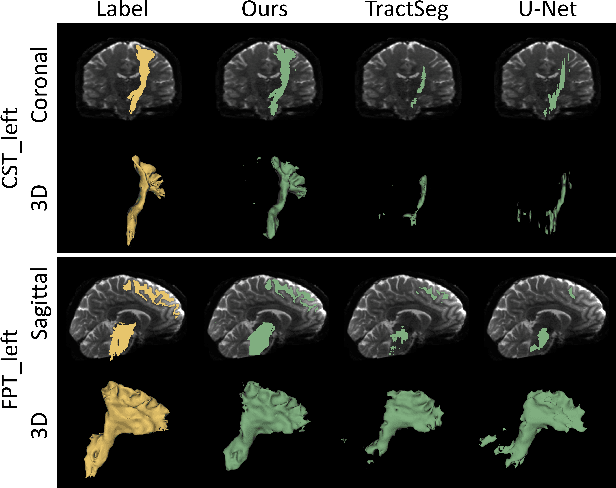
White matter (WM) tract segmentation based on diffusion magnetic resonance imaging (dMRI) plays an important role in the analysis of human health and brain diseases. However, the annotation of WM tracts is time-consuming and needs experienced neuroanatomists. In this study, to explore tract segmentation in the challenging setting of minimal annotations, we propose a novel framework utilizing only one annotated subject (subject-level one-shot) for tract segmentation. Our method is constructed by proposed registration-based peak augmentation (RPA) and uncertainty-based refining (URe) modules. RPA module synthesizes pseudo subjects and their corresponding labels to improve the tract segmentation performance. The proposed URe module alleviates the negative influence of the low-confidence voxels on pseudo subjects. Experimental results show that our method outperforms other state-of-the-art methods by a large margin, and our proposed modules are effective. Overall, our method achieves accurate whole-brain tract segmentation with only one annotated subject. Our code is available at https://github.com/HaoXu0507/ISBI2023-One-Shot-WM-Tract-Segmentation.
TractoFormer: A Novel Fiber-level Whole Brain Tractography Analysis Framework Using Spectral Embedding and Vision Transformers
Jul 11, 2022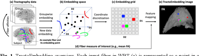
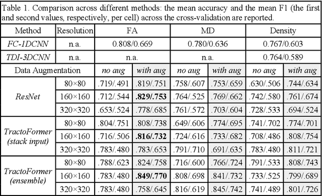
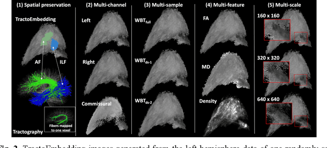
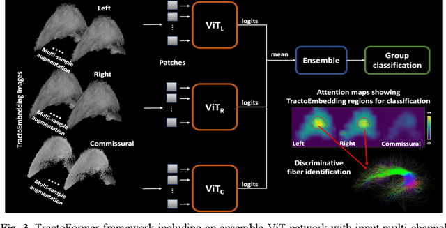
Diffusion MRI tractography is an advanced imaging technique for quantitative mapping of the brain's structural connectivity. Whole brain tractography (WBT) data contains over hundreds of thousands of individual fiber streamlines (estimated brain connections), and this data is usually parcellated to create compact representations for data analysis applications such as disease classification. In this paper, we propose a novel parcellation-free WBT analysis framework, TractoFormer, that leverages tractography information at the level of individual fiber streamlines and provides a natural mechanism for interpretation of results using the attention mechanism of transformers. TractoFormer includes two main contributions. First, we propose a novel and simple 2D image representation of WBT, TractoEmbedding, to encode 3D fiber spatial relationships and any feature of interest that can be computed from individual fibers (such as FA or MD). Second, we design a network based on vision transformers (ViTs) that includes: 1) data augmentation to overcome model overfitting on small datasets, 2) identification of discriminative fibers for interpretation of results, and 3) ensemble learning to leverage fiber information from different brain regions. In a synthetic data experiment, TractoFormer successfully identifies discriminative fibers with simulated group differences. In a disease classification experiment comparing several methods, TractoFormer achieves the highest accuracy in classifying schizophrenia vs control. Discriminative fibers are identified in left hemispheric frontal and parietal superficial white matter regions, which have previously been shown to be affected in schizophrenia patients.