Feiyan Li
Rethinking Normalization Strategies and Convolutional Kernels for Multimodal Image Fusion
Nov 15, 2024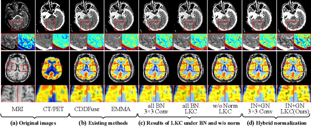

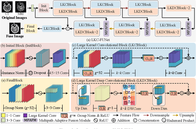
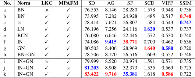
Abstract:Multimodal image fusion (MMIF) aims to integrate information from different modalities to obtain a comprehensive image, aiding downstream tasks. However, existing methods tend to prioritize natural image fusion and focus on information complementary and network training strategies. They ignore the essential distinction between natural and medical image fusion and the influence of underlying components. This paper dissects the significant differences between the two tasks regarding fusion goals, statistical properties, and data distribution. Based on this, we rethink the suitability of the normalization strategy and convolutional kernels for end-to-end MMIF.Specifically, this paper proposes a mixture of instance normalization and group normalization to preserve sample independence and reinforce intrinsic feature correlation.This strategy promotes the potential of enriching feature maps, thus boosting fusion performance. To this end, we further introduce the large kernel convolution, effectively expanding receptive fields and enhancing the preservation of image detail. Moreover, the proposed multipath adaptive fusion module recalibrates the decoder input with features of various scales and receptive fields, ensuring the transmission of crucial information. Extensive experiments demonstrate that our method exhibits state-of-the-art performance in multiple fusion tasks and significantly improves downstream applications. The code is available at https://github.com/HeDan-11/LKC-FUNet.
MyoPS: A Benchmark of Myocardial Pathology Segmentation Combining Three-Sequence Cardiac Magnetic Resonance Images
Jan 10, 2022
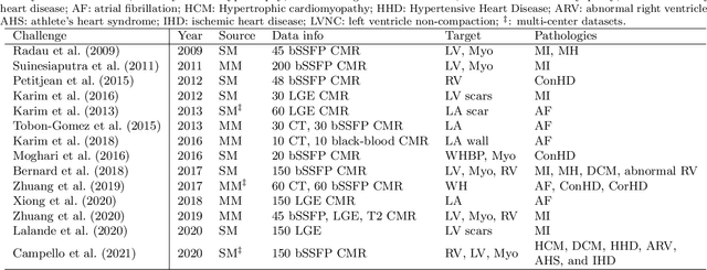
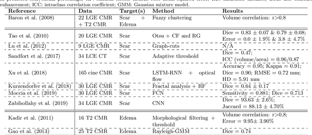
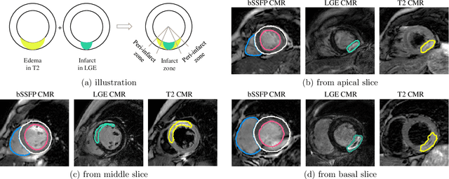
Abstract:Assessment of myocardial viability is essential in diagnosis and treatment management of patients suffering from myocardial infarction, and classification of pathology on myocardium is the key to this assessment. This work defines a new task of medical image analysis, i.e., to perform myocardial pathology segmentation (MyoPS) combining three-sequence cardiac magnetic resonance (CMR) images, which was first proposed in the MyoPS challenge, in conjunction with MICCAI 2020. The challenge provided 45 paired and pre-aligned CMR images, allowing algorithms to combine the complementary information from the three CMR sequences for pathology segmentation. In this article, we provide details of the challenge, survey the works from fifteen participants and interpret their methods according to five aspects, i.e., preprocessing, data augmentation, learning strategy, model architecture and post-processing. In addition, we analyze the results with respect to different factors, in order to examine the key obstacles and explore potential of solutions, as well as to provide a benchmark for future research. We conclude that while promising results have been reported, the research is still in the early stage, and more in-depth exploration is needed before a successful application to the clinics. Note that MyoPS data and evaluation tool continue to be publicly available upon registration via its homepage (www.sdspeople.fudan.edu.cn/zhuangxiahai/0/myops20/).
 Add to Chrome
 Add to Firefox
 Add to Edge