Adam S. Wang
Unsupervised Training of a Dynamic Context-Aware Deep Denoising Framework for Low-Dose Fluoroscopic Imaging
Oct 29, 2024


Abstract:Fluoroscopy is critical for real-time X-ray visualization in medical imaging. However, low-dose images are compromised by noise, potentially affecting diagnostic accuracy. Noise reduction is crucial for maintaining image quality, especially given such challenges as motion artifacts and the limited availability of clean data in medical imaging. To address these issues, we propose an unsupervised training framework for dynamic context-aware denoising of fluoroscopy image sequences. First, we train the multi-scale recurrent attention U-Net (MSR2AU-Net) without requiring clean data to address the initial noise. Second, we incorporate a knowledge distillation-based uncorrelated noise suppression module and a recursive filtering-based correlated noise suppression module enhanced with motion compensation to further improve motion compensation and achieve superior denoising performance. Finally, we introduce a novel approach by combining these modules with a pixel-wise dynamic object motion cross-fusion matrix, designed to adapt to motion, and an edge-preserving loss for precise detail retention. To validate the proposed method, we conducted extensive numerical experiments on medical image datasets, including 3500 fluoroscopy images from dynamic phantoms (2,400 images for training, 1,100 for testing) and 350 clinical images from a spinal surgery patient. Moreover, we demonstrated the robustness of our approach across different imaging modalities by testing it on the publicly available 2016 Low Dose CT Grand Challenge dataset, using 4,800 images for training and 1,136 for testing. The results demonstrate that the proposed approach outperforms state-of-the-art unsupervised algorithms in both visual quality and quantitative evaluation while achieving comparable performance to well-established supervised learning methods across low-dose fluoroscopy and CT imaging.
An Analysis of Scatter Characteristics in X-ray CT Spectral Correction
Dec 30, 2020


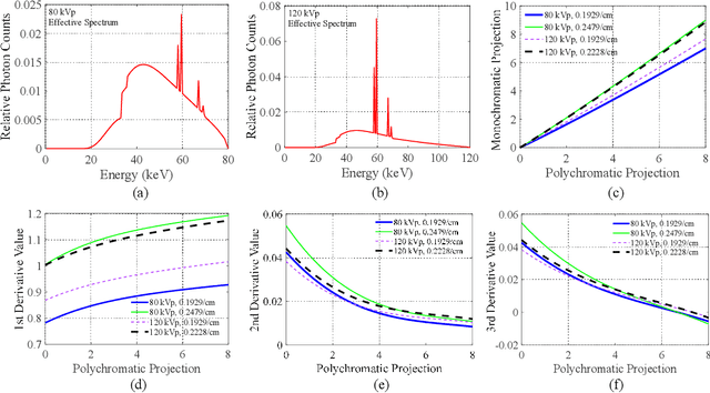
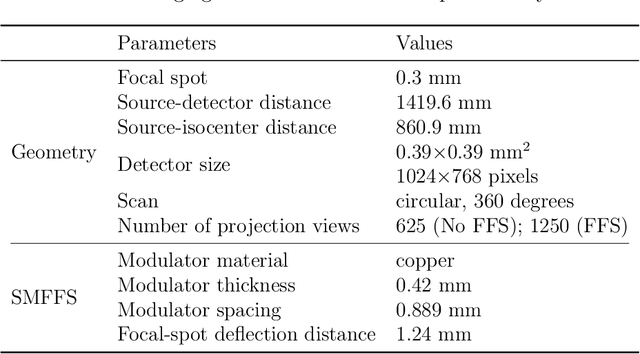
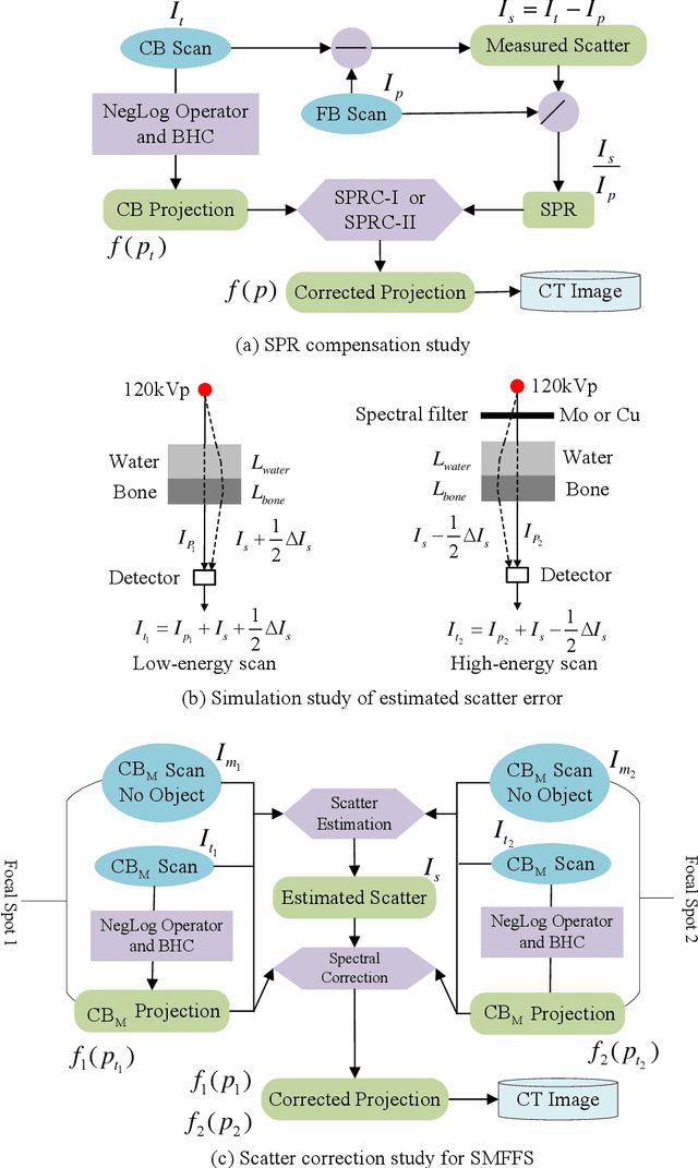
Abstract:X-ray scatter remains a major physics challenge in volumetric computed tomography (CT), whose physical and statistical behaviors have been commonly leveraged in order to eliminate its impact on CT image quality. In this work, we conduct an in-depth derivation of how the scatter distribution and scatter to primary ratio (SPR) will change during the spectral correction, leading to an interesting finding on the property of scatter: when applying the spectral correction before scatter is removed, the impact of SPR on a CT projection will be scaled by the first derivative of the mapping function; while the scatter distribution in the transmission domain will be scaled by the product of the first derivative of the mapping function and a natural exponential of the projection difference before and after the mapping. Such a characterization of scatter's behavior provides an analytic approach of compensating for the SPR as well as approximating the change of scatter distribution after spectral correction, even though both of them might be significantly distorted as the linearization mapping function in spectral correction could vary a lot from one detector pixel to another. We conduct an evaluation of SPR compensations on a Catphan phantom and an anthropomorphic chest phantom to validate the characteristics of scatter. In addition, this scatter property is also directly adopted into CT imaging using a spectral modulator with flying focal spot technology (SMFFS) as an example to demonstrate its potential in practical applications.
Assessing Robustness to Noise: Low-Cost Head CT Triage
Mar 29, 2020


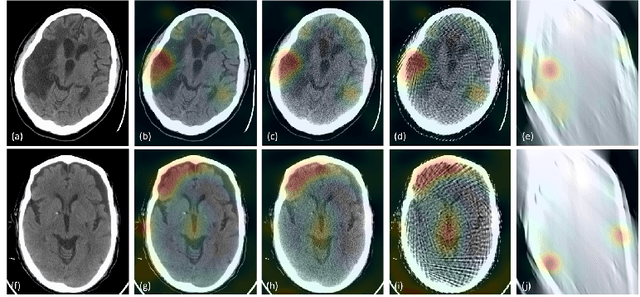
Abstract:Automated medical image classification with convolutional neural networks (CNNs) has great potential to impact healthcare, particularly in resource-constrained healthcare systems where fewer trained radiologists are available. However, little is known about how well a trained CNN can perform on images with the increased noise levels, different acquisition protocols, or additional artifacts that may arise when using low-cost scanners, which can be underrepresented in datasets collected from well-funded hospitals. In this work, we investigate how a model trained to triage head computed tomography (CT) scans performs on images acquired with reduced x-ray tube current, fewer projections per gantry rotation, and limited angle scans. These changes can reduce the cost of the scanner and demands on electrical power but come at the expense of increased image noise and artifacts. We first develop a model to triage head CTs and report an area under the receiver operating characteristic curve (AUROC) of 0.77. We then show that the trained model is robust to reduced tube current and fewer projections, with the AUROC dropping only 0.65% for images acquired with a 16x reduction in tube current and 0.22% for images acquired with 8x fewer projections. Finally, for significantly degraded images acquired by a limited angle scan, we show that a model trained specifically to classify such images can overcome the technological limitations to reconstruction and maintain an AUROC within 0.09% of the original model.
 Add to Chrome
 Add to Firefox
 Add to Edge