Assessing Robustness to Noise: Low-Cost Head CT Triage
Paper and Code
Mar 29, 2020


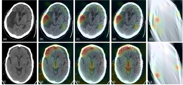
Automated medical image classification with convolutional neural networks (CNNs) has great potential to impact healthcare, particularly in resource-constrained healthcare systems where fewer trained radiologists are available. However, little is known about how well a trained CNN can perform on images with the increased noise levels, different acquisition protocols, or additional artifacts that may arise when using low-cost scanners, which can be underrepresented in datasets collected from well-funded hospitals. In this work, we investigate how a model trained to triage head computed tomography (CT) scans performs on images acquired with reduced x-ray tube current, fewer projections per gantry rotation, and limited angle scans. These changes can reduce the cost of the scanner and demands on electrical power but come at the expense of increased image noise and artifacts. We first develop a model to triage head CTs and report an area under the receiver operating characteristic curve (AUROC) of 0.77. We then show that the trained model is robust to reduced tube current and fewer projections, with the AUROC dropping only 0.65% for images acquired with a 16x reduction in tube current and 0.22% for images acquired with 8x fewer projections. Finally, for significantly degraded images acquired by a limited angle scan, we show that a model trained specifically to classify such images can overcome the technological limitations to reconstruction and maintain an AUROC within 0.09% of the original model.
 Add to Chrome
 Add to Firefox
 Add to Edge