Xinzi Sun
A Knowledge-Based Decision Support System for In Vitro Fertilization Treatment
Jan 27, 2022
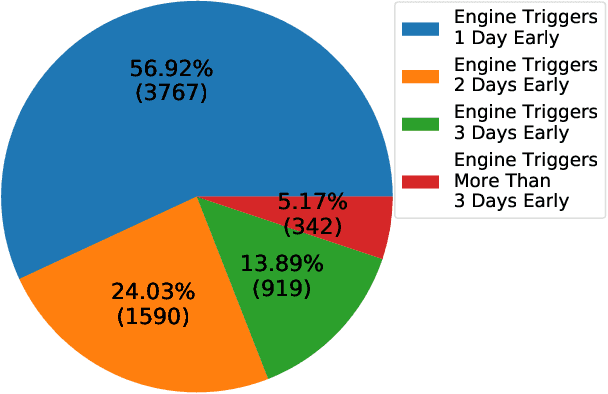
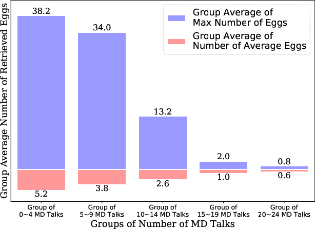
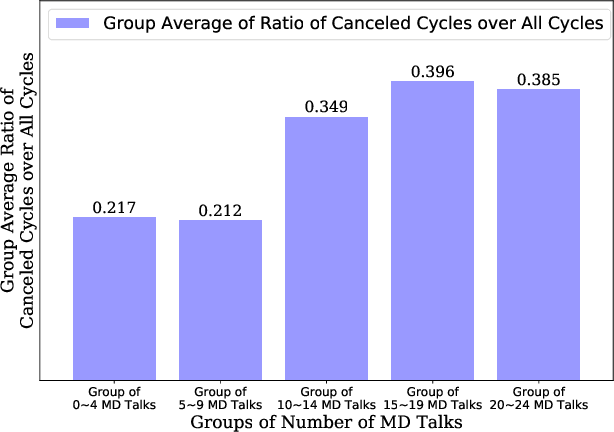
Abstract:In Vitro Fertilization (IVF) is the most widely used Assisted Reproductive Technology (ART). IVF usually involves controlled ovarian stimulation, oocyte retrieval, fertilization in the laboratory with subsequent embryo transfer. The first two steps correspond with follicular phase of females and ovulation in their menstrual cycle. Therefore, we refer to it as the treatment cycle in our paper. The treatment cycle is crucial because the stimulation medications in IVF treatment are applied directly on patients. In order to optimize the stimulation effects and lower the side effects of the stimulation medications, prompt treatment adjustments are in need. In addition, the quality and quantity of the retrieved oocytes have a significant effect on the outcome of the following procedures. To improve the IVF success rate, we propose a knowledge-based decision support system that can provide medical advice on the treatment protocol and medication adjustment for each patient visit during IVF treatment cycle. Our system is efficient in data processing and light-weighted which can be easily embedded into electronic medical record systems. Moreover, an oocyte retrieval oriented evaluation demonstrates that our system performs well in terms of accuracy of advice for the protocols and medications.
Colorectal Polyp Detection in Real-world Scenario: Design and Experiment Study
Jan 11, 2021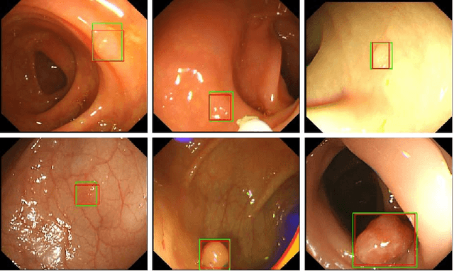
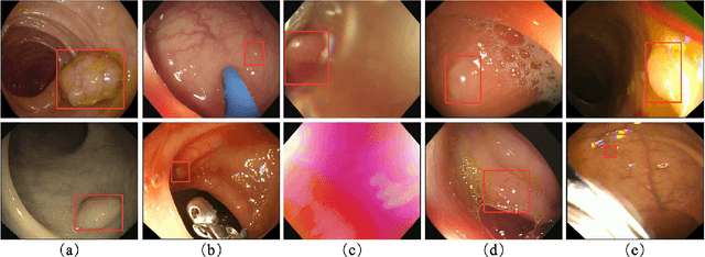


Abstract:Colorectal polyps are abnormal tissues growing on the intima of the colon or rectum with a high risk of developing into colorectal cancer, the third leading cause of cancer death worldwide. Early detection and removal of colon polyps via colonoscopy have proved to be an effective approach to prevent colorectal cancer. Recently, various CNN-based computer-aided systems have been developed to help physicians detect polyps. However, these systems do not perform well in real-world colonoscopy operations due to the significant difference between images in a real colonoscopy and those in the public datasets. Unlike the well-chosen clear images with obvious polyps in the public datasets, images from a colonoscopy are often blurry and contain various artifacts such as fluid, debris, bubbles, reflection, specularity, contrast, saturation, and medical instruments, with a wide variety of polyps of different sizes, shapes, and textures. All these factors pose a significant challenge to effective polyp detection in a colonoscopy. To this end, we collect a private dataset that contains 7,313 images from 224 complete colonoscopy procedures. This dataset represents realistic operation scenarios and thus can be used to better train the models and evaluate a system's performance in practice. We propose an integrated system architecture to address the unique challenges for polyp detection. Extensive experiments results show that our system can effectively detect polyps in a colonoscopy with excellent performance in real time.
Colorectal Polyp Segmentation by U-Net with Dilation Convolution
Dec 26, 2019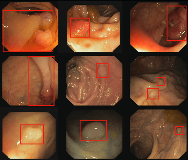

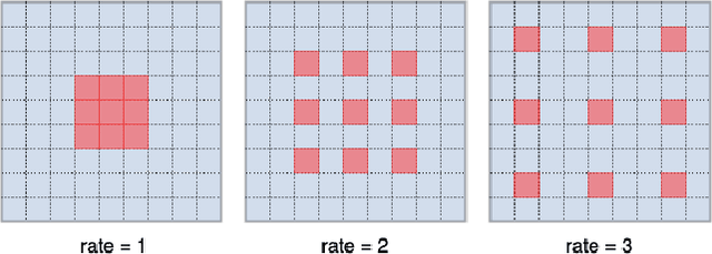

Abstract:Colorectal cancer (CRC) is one of the most commonly diagnosed cancers and a leading cause of cancer deaths in the United States. Colorectal polyps that grow on the intima of the colon or rectum is an important precursor for CRC. Currently, the most common way for colorectal polyp detection and precancerous pathology is the colonoscopy. Therefore, accurate colorectal polyp segmentation during the colonoscopy procedure has great clinical significance in CRC early detection and prevention. In this paper, we propose a novel end-to-end deep learning framework for the colorectal polyp segmentation. The model we design consists of an encoder to extract multi-scale semantic features and a decoder to expand the feature maps to a polyp segmentation map. We improve the feature representation ability of the encoder by introducing the dilated convolution to learn high-level semantic features without resolution reduction. We further design a simplified decoder which combines multi-scale semantic features with fewer parameters than the traditional architecture. Furthermore, we apply three post processing techniques on the output segmentation map to improve colorectal polyp detection performance. Our method achieves state-of-the-art results on CVC-ClinicDB and ETIS-Larib Polyp DB.
Mini Lesions Detection on Diabetic Retinopathy Images via Large Scale CNN Features
Nov 19, 2019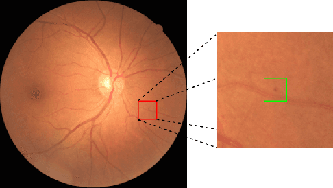
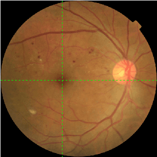
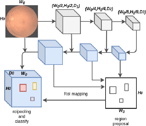
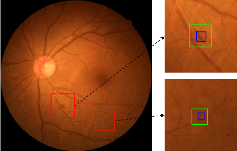
Abstract:Diabetic retinopathy (DR) is a diabetes complication that affects eyes. DR is a primary cause of blindness in working-age people and it is estimated that 3 to 4 million people with diabetes are blinded by DR every year worldwide. Early diagnosis have been considered an effective way to mitigate such problem. The ultimate goal of our research is to develop novel machine learning techniques to analyze the DR images generated by the fundus camera for automatically DR diagnosis. In this paper, we focus on identifying small lesions on DR fundus images. The results from our analysis, which include the lesion category and their exact locations in the image, can be used to facilitate the determination of DR severity (indicated by DR stages). Different from traditional object detection for natural images, lesion detection for fundus images have unique challenges. Specifically, the size of a lesion instance is usually very small, compared with the original resolution of the fundus images, making them diffcult to be detected. We analyze the lesion-vs-image scale carefully and propose a large-size feature pyramid network (LFPN) to preserve more image details for mini lesion instance detection. Our method includes an effective region proposal strategy to increase the sensitivity. The experimental results show that our proposed method is superior to the original feature pyramid network (FPN) method and Faster RCNN.
AFP-Net: Realtime Anchor-Free Polyp Detection in Colonoscopy
Sep 26, 2019


Abstract:Colorectal cancer (CRC) is a common and lethal disease. Globally, CRC is the third most commonly diagnosed cancer in males and the second in females. For colorectal cancer, the best screening test available is the colonoscopy. During a colonoscopic procedure, a tiny camera at the tip of the endoscope generates a video of the internal mucosa of the colon. The video data are displayed on a monitor for the physician to examine the lining of the entire colon and check for colorectal polyps. Detection and removal of colorectal polyps are associated with a reduction in mortality from colorectal cancer. However, the miss rate of polyp detection during colonoscopy procedure is often high even for very experienced physicians. The reason lies in the high variation of polyp in terms of shape, size, textural, color and illumination. Though challenging, with the great advances in object detection techniques, automated polyp detection still demonstrates a great potential in reducing the false negative rate while maintaining a high precision. In this paper, we propose a novel anchor free polyp detector that can localize polyps without using predefined anchor boxes. To further strengthen the model, we leverage a Context Enhancement Module and Cosine Ground truth Projection. Our approach can respond in real time while achieving state-of-the-art performance with 99.36% precision and 96.44% recall.
3D Anchor-Free Lesion Detector on Computed Tomography Scans
Aug 29, 2019


Abstract:Lesions are injuries and abnormal tissues in the human body. Detecting lesions in 3D Computed Tomography (CT) scans can be time-consuming even for very experienced physicians and radiologists. In recent years, CNN based lesion detectors have demonstrated huge potentials. Most of current state-of-the-art lesion detectors employ anchors to enumerate all possible bounding boxes with respect to the dataset in process. This anchor mechanism greatly improves the detection performance while also constraining the generalization ability of detectors. In this paper, we propose an anchor-free lesion detector. The anchor mechanism is removed and lesions are formalized as single keypoints. By doing so, we witness a considerable performance gain in terms of both accuracy and inference speed compared with the anchor-based baseline
 Add to Chrome
 Add to Firefox
 Add to Edge