Qilei Chen
Retinopathy of Prematurity Stage Diagnosis Using Object Segmentation and Convolutional Neural Networks
Apr 03, 2020
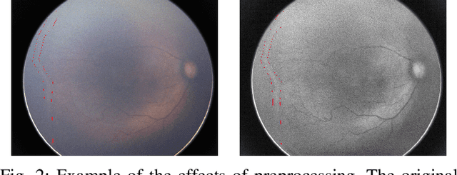

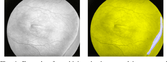
Abstract:Retinopathy of Prematurity (ROP) is an eye disorder primarily affecting premature infants with lower weights. It causes proliferation of vessels in the retina and could result in vision loss and, eventually, retinal detachment, leading to blindness. While human experts can easily identify severe stages of ROP, the diagnosis of earlier stages, which are the most relevant to determining treatment choice, are much more affected by variability in subjective interpretations of human experts. In recent years, there has been a significant effort to automate the diagnosis using deep learning. This paper builds upon the success of previous models and develops a novel architecture, which combines object segmentation and convolutional neural networks (CNN) to construct an effective classifier of ROP stages 1-3 based on neonatal retinal images. Motivated by the fact that the formation and shape of a demarcation line in the retina is the distinguishing feature between earlier ROP stages, our proposed system first trains an object segmentation model to identify the demarcation line at a pixel level and adds the resulting mask as an additional "color" channel in the original image. Then, the system trains a CNN classifier based on the processed images to leverage information from both the original image and the mask, which helps direct the model's attention to the demarcation line. In a number of careful experiments comparing its performance to previous object segmentation systems and CNN-only systems trained on our dataset, our novel architecture significantly outperforms previous systems in accuracy, demonstrating the effectiveness of our proposed pipeline.
Pseudo-Labeling for Small Lesion Detection on Diabetic Retinopathy Images
Mar 26, 2020


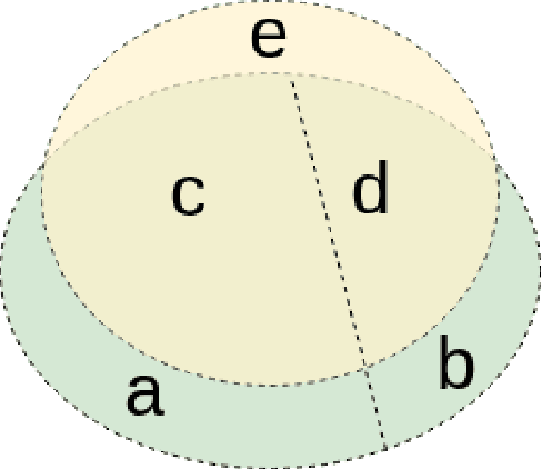
Abstract:Diabetic retinopathy (DR) is a primary cause of blindness in working-age people worldwide. About 3 to 4 million people with diabetes become blind because of DR every year. Diagnosis of DR through color fundus images is a common approach to mitigate such problem. However, DR diagnosis is a difficult and time consuming task, which requires experienced clinicians to identify the presence and significance of many small features on high resolution images. Convolutional Neural Network (CNN) has proved to be a promising approach for automatic biomedical image analysis recently. In this work, we investigate lesion detection on DR fundus images with CNN-based object detection methods. Lesion detection on fundus images faces two unique challenges. The first one is that our dataset is not fully labeled, i.e., only a subset of all lesion instances are marked. Not only will these unlabeled lesion instances not contribute to the training of the model, but also they will be mistakenly counted as false negatives, leading the model move to the opposite direction. The second challenge is that the lesion instances are usually very small, making them difficult to be found by normal object detectors. To address the first challenge, we introduce an iterative training algorithm for the semi-supervised method of pseudo-labeling, in which a considerable number of unlabeled lesion instances can be discovered to boost the performance of the lesion detector. For the small size targets problem, we extend both the input size and the depth of feature pyramid network (FPN) to produce a large CNN feature map, which can preserve the detail of small lesions and thus enhance the effectiveness of the lesion detector. The experimental results show that our proposed methods significantly outperform the baselines.
Mini Lesions Detection on Diabetic Retinopathy Images via Large Scale CNN Features
Nov 19, 2019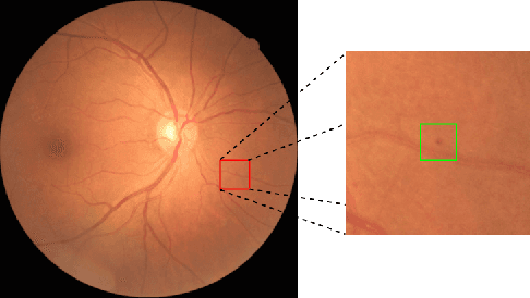
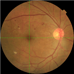
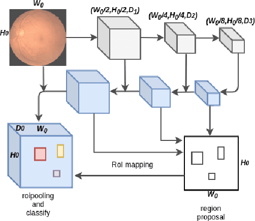
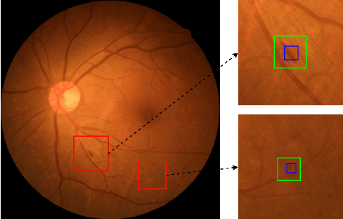
Abstract:Diabetic retinopathy (DR) is a diabetes complication that affects eyes. DR is a primary cause of blindness in working-age people and it is estimated that 3 to 4 million people with diabetes are blinded by DR every year worldwide. Early diagnosis have been considered an effective way to mitigate such problem. The ultimate goal of our research is to develop novel machine learning techniques to analyze the DR images generated by the fundus camera for automatically DR diagnosis. In this paper, we focus on identifying small lesions on DR fundus images. The results from our analysis, which include the lesion category and their exact locations in the image, can be used to facilitate the determination of DR severity (indicated by DR stages). Different from traditional object detection for natural images, lesion detection for fundus images have unique challenges. Specifically, the size of a lesion instance is usually very small, compared with the original resolution of the fundus images, making them diffcult to be detected. We analyze the lesion-vs-image scale carefully and propose a large-size feature pyramid network (LFPN) to preserve more image details for mini lesion instance detection. Our method includes an effective region proposal strategy to increase the sensitivity. The experimental results show that our proposed method is superior to the original feature pyramid network (FPN) method and Faster RCNN.
 Add to Chrome
 Add to Firefox
 Add to Edge