Thomas Kirchner
Machine learning enabled multiple illumination quantitative optoacoustic imaging of blood oxygenation in humans
Feb 03, 2022


Abstract:Optoacoustic (OA) imaging is a promising modality for quantifying blood oxygen saturation (sO$_2$) in various biomedical applications - in diagnosis, monitoring of organ function or even tumor treatment planning. We present an accurate and practically feasible real-time capable method for quantitative imaging of sO$_2$ based on combining multispectral (MS) and multiple illumination (MI) OA imaging with learned spectral decoloring (LSD). For this purpose we developed a hybrid real-time MI MS OA imaging setup with ultrasound (US) imaging capability; we trained gradient boosting machines on MI spectrally colored absorbed energy spectra generated by generic Monte Carlo simulations, and used the trained models to estimate sO$_2$ on real OA measurements. We validated MI-LSD in silico and on in vivo image sequences of radial arteries and accompanying veins of five healthy human volunteers. We compared the performance of the method to prior LSD work and conventional linear unmixing. MI-LSD provided highly accurate results in silico and consistently plausible results in vivo. This preliminary study shows a potentially high applicability of quantitative OA oximetry imaging, using our method.
Quantitative photoacoustic oximetry imaging by multiple illumination learned spectral decoloring
Feb 22, 2021


Abstract:Significance: Quantitative measurement of blood oxygen saturation (sO$_2$) with photoacoustic (PA) imaging is one of the most sought after goals of quantitative PA imaging research due to its wide range of biomedical applications. Aim: A method for accurate and applicable real-time quantification of local sO$_2$ with PA imaging. Approach: We combine multiple illumination (MI) sensing with learned spectral decoloring (LSD); training on Monte Carlo simulations of spectrally colored absorbed energy spectra, in order to apply the trained models to real PA measurements. We validate our combined MI-LSD method on a highly reliable, reproducible and easily scalable phantom model, based on copper and nickel sulfate solutions. Results: With this sulfate model we see a consistently high estimation accuracy using MI-LSD, with median absolute estimation errors of 2.5 to 4.5 percentage points. We further find fewer outliers in MI-LSD estimates compared to LSD. Random forest regressors outperform previously reported neural network approaches. Conclusions: Random forest based MI-LSD is a promising method for accurate quantitative PA oximetry imaging.
Uncertainty-aware performance assessment of optical imaging modalities with invertible neural networks
Mar 08, 2019


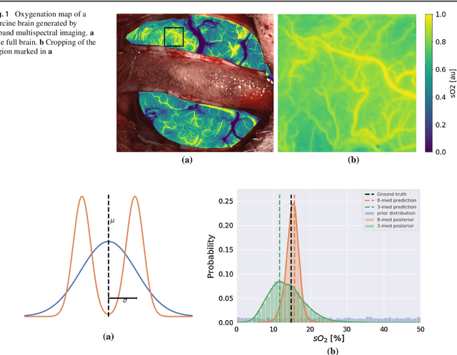
Abstract:Purpose: Optical imaging is evolving as a key technique for advanced sensing in the operating room. Recent research has shown that machine learning algorithms can be used to address the inverse problem of converting pixel-wise multispectral reflectance measurements to underlying tissue parameters, such as oxygenation. Assessment of the specific hardware used in conjunction with such algorithms, however, has not properly addressed the possibility that the problem may be ill-posed. Methods: We present a novel approach to the assessment of optical imaging modalities, which is sensitive to the different types of uncertainties that may occur when inferring tissue parameters. Based on the concept of invertible neural networks, our framework goes beyond point estimates and maps each multispectral measurement to a full posterior probability distribution which is capable of representing ambiguity in the solution via multiple modes. Performance metrics for a hardware setup can then be computed from the characteristics of the posteriors. Results: Application of the assessment framework to the specific use case of camera selection for physiological parameter estimation yields the following insights: (1) Estimation of tissue oxygenation from multispectral images is a well-posed problem, while (2) blood volume fraction may not be recovered without ambiguity. (3) In general, ambiguity may be reduced by increasing the number of spectral bands in the camera. Conclusion: Our method could help to optimize optical camera design in an application-specific manner.
Estimation of blood oxygenation with learned spectral decoloring for quantitative photoacoustic imaging (LSD-qPAI)
Feb 15, 2019


Abstract:One of the main applications of photoacoustic (PA) imaging is the recovery of functional tissue properties, such as blood oxygenation (sO2). This is typically achieved by linear spectral unmixing of relevant chromophores from multispectral photoacoustic images. Despite the progress that has been made towards quantitative PA imaging (qPAI), most sO2 estimation methods yield poor results in realistic settings. In this work, we tackle the challenge by employing learned spectral decoloring for quantitative photoacoustic imaging (LSD-qPAI) to obtain quantitative estimates for blood oxygenation. LSD-qPAI computes sO2 directly from pixel-wise initial pressure spectra Sp0, which are vectors comprised of the initial pressure at the same spatial location over all recorded wavelengths. Initial results suggest that LSD-qPAI is able to obtain accurate sO2 estimates directly from multispectral photoacoustic measurements in silico and plausible estimates in vivo.
Context encoding enables machine learning-based quantitative photoacoustics
Jan 29, 2018Abstract:Real-time monitoring of functional tissue parameters, such as local blood oxygenation, based on optical imaging could provide groundbreaking advances in the diagnosis and interventional therapy of various diseases. While photoacoustic (PA) imaging is a novel modality with great potential to measure optical absorption deep inside tissue, quantification of the measurements remains a major challenge. In this paper, we introduce the first machine learning based approach to quantitative PA imaging (qPAI), which relies on learning the fluence in a voxel to deduce the corresponding optical absorption. The method encodes relevant information of the measured signal and the characteristics of the imaging system in voxel-based feature vectors, which allow the generation of thousands of training samples from a single simulated PA image. Comprehensive in silico experiments suggest that context encoding (CE)-qPAI enables highly accurate and robust quantification of the local fluence and thereby the optical absorption from PA images.
 Add to Chrome
 Add to Firefox
 Add to Edge