 Add to Chrome
 Add to Firefox
 Add to Edge
SGW-GAN: Sliced Gromov-Wasserstein Guided GANs for Retinal Fundus Image Enhancement
Jan 19, 2026Retinal fundus photography is indispensable for ophthalmic screening and diagnosis, yet image quality is often degraded by noise, artifacts, and uneven illumination. Recent GAN- and diffusion-based enhancement methods improve perceptual quality by aligning degraded images with high-quality distributions, but our analysis shows that this focus can distort intra-class geometry: clinically related samples become dispersed, disease-class boundaries blur, and downstream tasks such as grading or lesion detection are harmed. The Gromov Wasserstein (GW) discrepancy offers a principled solution by aligning distributions through internal pairwise distances, naturally preserving intra-class structure, but its high computational cost restricts practical use. To overcome this, we propose SGW-GAN, the first framework to incorporate Sliced GW (SGW) into retinal image enhancement. SGW approximates GW via random projections, retaining relational fidelity while greatly reducing cost. Experiments on public datasets show that SGW-GAN produces visually compelling enhancements, achieves superior diabetic retinopathy grading, and reports the lowest GW discrepancy across disease labels, demonstrating both efficiency and clinical fidelity for unpaired medical image enhancement.
RetinalGPT: A Retinal Clinical Preference Conversational Assistant Powered by Large Vision-Language Models
Mar 06, 2025Recently, Multimodal Large Language Models (MLLMs) have gained significant attention for their remarkable ability to process and analyze non-textual data, such as images, videos, and audio. Notably, several adaptations of general-domain MLLMs to the medical field have been explored, including LLaVA-Med. However, these medical adaptations remain insufficiently advanced in understanding and interpreting retinal images. In contrast, medical experts emphasize the importance of quantitative analyses for disease detection and interpretation. This underscores a gap between general-domain and medical-domain MLLMs: while general-domain MLLMs excel in broad applications, they lack the specialized knowledge necessary for precise diagnostic and interpretative tasks in the medical field. To address these challenges, we introduce \textit{RetinalGPT}, a multimodal conversational assistant for clinically preferred quantitative analysis of retinal images. Specifically, we achieve this by compiling a large retinal image dataset, developing a novel data pipeline, and employing customized visual instruction tuning to enhance both retinal analysis and enrich medical knowledge. In particular, RetinalGPT outperforms MLLM in the generic domain by a large margin in the diagnosis of retinal diseases in 8 benchmark retinal datasets. Beyond disease diagnosis, RetinalGPT features quantitative analyses and lesion localization, representing a pioneering step in leveraging LLMs for an interpretable and end-to-end clinical research framework. The code is available at https://github.com/Retinal-Research/RetinalGPT
EyeBench: A Call for More Rigorous Evaluation of Retinal Image Enhancement
Feb 20, 2025


Over the past decade, generative models have achieved significant success in enhancement fundus images.However, the evaluation of these models still presents a considerable challenge. A comprehensive evaluation benchmark for fundus image enhancement is indispensable for three main reasons: 1) The existing denoising metrics (e.g., PSNR, SSIM) are hardly to extend to downstream real-world clinical research (e.g., Vessel morphology consistency). 2) There is a lack of comprehensive evaluation for both paired and unpaired enhancement methods, along with the need for expert protocols to accurately assess clinical value. 3) An ideal evaluation system should provide insights to inform future developments of fundus image enhancement. To this end, we propose a novel comprehensive benchmark, EyeBench, to provide insights that align enhancement models with clinical needs, offering a foundation for future work to improve the clinical relevance and applicability of generative models for fundus image enhancement. EyeBench has three appealing properties: 1) multi-dimensional clinical alignment downstream evaluation: In addition to evaluating the enhancement task, we provide several clinically significant downstream tasks for fundus images, including vessel segmentation, DR grading, denoising generalization, and lesion segmentation. 2) Medical expert-guided evaluation design: We introduce a novel dataset that promote comprehensive and fair comparisons between paired and unpaired methods and includes a manual evaluation protocol by medical experts. 3) Valuable insights: Our benchmark study provides a comprehensive and rigorous evaluation of existing methods across different downstream tasks, assisting medical experts in making informed choices. Additionally, we offer further analysis of the challenges faced by existing methods. The code is available at \url{https://github.com/Retinal-Research/EyeBench}
Context-Aware Optimal Transport Learning for Retinal Fundus Image Enhancement
Sep 12, 2024


Retinal fundus photography offers a non-invasive way to diagnose and monitor a variety of retinal diseases, but is prone to inherent quality glitches arising from systemic imperfections or operator/patient-related factors. However, high-quality retinal images are crucial for carrying out accurate diagnoses and automated analyses. The fundus image enhancement is typically formulated as a distribution alignment problem, by finding a one-to-one mapping between a low-quality image and its high-quality counterpart. This paper proposes a context-informed optimal transport (OT) learning framework for tackling unpaired fundus image enhancement. In contrast to standard generative image enhancement methods, which struggle with handling contextual information (e.g., over-tampered local structures and unwanted artifacts), the proposed context-aware OT learning paradigm better preserves local structures and minimizes unwanted artifacts. Leveraging deep contextual features, we derive the proposed context-aware OT using the earth mover's distance and show that the proposed context-OT has a solid theoretical guarantee. Experimental results on a large-scale dataset demonstrate the superiority of the proposed method over several state-of-the-art supervised and unsupervised methods in terms of signal-to-noise ratio, structural similarity index, as well as two downstream tasks. The code is available at \url{https://github.com/Retinal-Research/Contextual-OT}.
RBAD: A Dataset and Benchmark for Retinal Vessels Branching Angle Detection
Jul 17, 2024

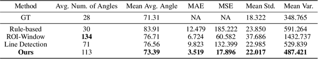

Detecting retinal image analysis, particularly the geometrical features of branching points, plays an essential role in diagnosing eye diseases. However, existing methods used for this purpose often are coarse-level and lack fine-grained analysis for efficient annotation. To mitigate these issues, this paper proposes a novel method for detecting retinal branching angles using a self-configured image processing technique. Additionally, we offer an open-source annotation tool and a benchmark dataset comprising 40 images annotated with retinal branching angles. Our methodology for retinal branching angle detection and calculation is detailed, followed by a benchmark analysis comparing our method with previous approaches. The results indicate that our method is robust under various conditions with high accuracy and efficiency, which offers a valuable instrument for ophthalmic research and clinical applications.