Nicolás Gaggion
ChronoRoot 2.0: An Open AI-Powered Platform for 2D Temporal Plant Phenotyping
Apr 20, 2025


Abstract:The analysis of plant developmental plasticity, including root system architecture, is fundamental to understanding plant adaptability and development, particularly in the context of climate change and agricultural sustainability. While significant advances have been made in plant phenotyping technologies, comprehensive temporal analysis of root development remains challenging, with most existing solutions providing either limited throughput or restricted structural analysis capabilities. Here, we present ChronoRoot 2.0, an integrated open-source platform that combines affordable hardware with advanced artificial intelligence to enable sophisticated temporal plant phenotyping. The system introduces several major advances, offering an integral perspective of seedling development: (i) simultaneous multi-organ tracking of six distinct plant structures, (ii) quality control through real-time validation, (iii) comprehensive architectural measurements including novel gravitropic response parameters, and (iv) dual specialized user interfaces for both architectural analysis and high-throughput screening. We demonstrate the system's capabilities through three use cases for Arabidopsis thaliana: characterization of circadian growth patterns under different light conditions, detailed analysis of gravitropic responses in transgenic plants, and high-throughput screening of etiolation responses across multiple genotypes. ChronoRoot 2.0 maintains its predecessor's advantages of low cost and modularity while significantly expanding its capabilities, making sophisticated temporal phenotyping more accessible to the broader plant science community. The system's open-source nature, combined with extensive documentation and containerized deployment options, ensures reproducibility and enables community-driven development of new analytical capabilities.
Fitting Skeletal Models via Graph-based Learning
Sep 09, 2024


Abstract:Skeletonization is a popular shape analysis technique that models an object's interior as opposed to just its boundary. Fitting template-based skeletal models is a time-consuming process requiring much manual parameter tuning. Recently, machine learning-based methods have shown promise for generating s-reps from object boundaries. In this work, we propose a new skeletonization method which leverages graph convolutional networks to produce skeletal representations (s-reps) from dense segmentation masks. The method is evaluated on both synthetic data and real hippocampus segmentations, achieving promising results and fast inference.
* This paper was presented at the 2024 IEEE International Symposium on Biomedical Imaging (ISBI)
Multi-view Hybrid Graph Convolutional Network for Volume-to-mesh Reconstruction in Cardiovascular MRI
Nov 22, 2023Abstract:Cardiovascular magnetic resonance imaging is emerging as a crucial tool to examine cardiac morphology and function. Essential to this endeavour are anatomical 3D surface and volumetric meshes derived from CMR images, which facilitate computational anatomy studies, biomarker discovery, and in-silico simulations. However, conventional surface mesh generation methods, such as active shape models and multi-atlas segmentation, are highly time-consuming and require complex processing pipelines to generate simulation-ready 3D meshes. In response, we introduce HybridVNet, a novel architecture for direct image-to-mesh extraction seamlessly integrating standard convolutional neural networks with graph convolutions, which we prove can efficiently handle surface and volumetric meshes by encoding them as graph structures. To further enhance accuracy, we propose a multiview HybridVNet architecture which processes both long axis and short axis CMR, showing that it can increase the performance of cardiac MR mesh generation. Our model combines traditional convolutional networks with variational graph generative models, deep supervision and mesh-specific regularisation. Experiments on a comprehensive dataset from the UK Biobank confirm the potential of HybridVNet to significantly advance cardiac imaging and computational cardiology by efficiently generating high-fidelity and simulation ready meshes from CMR images.
Unsupervised bias discovery in medical image segmentation
Sep 01, 2023Abstract:It has recently been shown that deep learning models for anatomical segmentation in medical images can exhibit biases against certain sub-populations defined in terms of protected attributes like sex or ethnicity. In this context, auditing fairness of deep segmentation models becomes crucial. However, such audit process generally requires access to ground-truth segmentation masks for the target population, which may not always be available, especially when going from development to deployment. Here we propose a new method to anticipate model biases in biomedical image segmentation in the absence of ground-truth annotations. Our unsupervised bias discovery method leverages the reverse classification accuracy framework to estimate segmentation quality. Through numerical experiments in synthetic and realistic scenarios we show how our method is able to successfully anticipate fairness issues in the absence of ground-truth labels, constituting a novel and valuable tool in this field.
CheXmask: a large-scale dataset of anatomical segmentation masks for multi-center chest x-ray images
Jul 06, 2023Abstract:The development of successful artificial intelligence models for chest X-ray analysis relies on large, diverse datasets with high-quality annotations. While several databases of chest X-ray images have been released, most include disease diagnosis labels but lack detailed pixel-level anatomical segmentation labels. To address this gap, we introduce an extensive chest X-ray multi-center segmentation dataset with uniform and fine-grain anatomical annotations for images coming from six well-known publicly available databases: CANDID-PTX, ChestX-ray8, Chexpert, MIMIC-CXR-JPG, Padchest, and VinDr-CXR, resulting in 676,803 segmentation masks. Our methodology utilizes the HybridGNet model to ensure consistent and high-quality segmentations across all datasets. Rigorous validation, including expert physician evaluation and automatic quality control, was conducted to validate the resulting masks. Additionally, we provide individualized quality indices per mask and an overall quality estimation per dataset. This dataset serves as a valuable resource for the broader scientific community, streamlining the development and assessment of innovative methodologies in chest X-ray analysis. The CheXmask dataset is publicly available at: \url{https://physionet.org/content/chexmask-cxr-segmentation-data/}.
Multi-center anatomical segmentation with heterogeneous labels via landmark-based models
Nov 14, 2022Abstract:Learning anatomical segmentation from heterogeneous labels in multi-center datasets is a common situation encountered in clinical scenarios, where certain anatomical structures are only annotated in images coming from particular medical centers, but not in the full database. Here we first show how state-of-the-art pixel-level segmentation models fail in naively learning this task due to domain memorization issues and conflicting labels. We then propose to adopt HybridGNet, a landmark-based segmentation model which learns the available anatomical structures using graph-based representations. By analyzing the latent space learned by both models, we show that HybridGNet naturally learns more domain-invariant feature representations, and provide empirical evidence in the context of chest X-ray multiclass segmentation. We hope these insights will shed light on the training of deep learning models with heterogeneous labels from public and multi-center datasets.
Improving anatomical plausibility in medical image segmentation via hybrid graph neural networks: applications to chest x-ray analysis
Apr 01, 2022


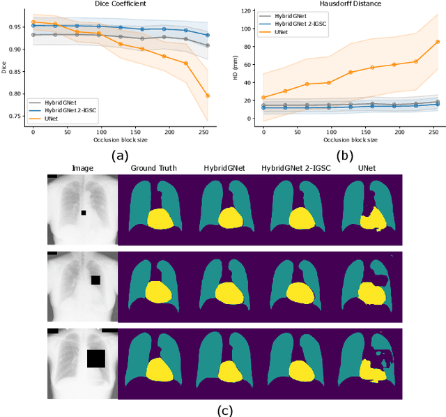
Abstract:Anatomical segmentation is a fundamental task in medical image computing, generally tackled with fully convolutional neural networks which produce dense segmentation masks. These models are often trained with loss functions such as cross-entropy or Dice, which assume pixels to be independent of each other, thus ignoring topological errors and anatomical inconsistencies. We address this limitation by moving from pixel-level to graph representations, which allow to naturally incorporate anatomical constraints by construction. To this end, we introduce HybridGNet, an encoder-decoder neural architecture that leverages standard convolutions for image feature encoding and graph convolutional neural networks (GCNNs) to decode plausible representations of anatomical structures. We also propose a novel image-to-graph skip connection layer which allows localized features to flow from standard convolutional blocks to GCNN blocks, and show that it improves segmentation accuracy. The proposed architecture is extensively evaluated in a variety of domain shift and image occlusion scenarios, and audited considering different types of demographic domain shift. Our comprehensive experimental setup compares HybridGNet with other landmark and pixel-based models for anatomical segmentation in chest x-ray images, and shows that it produces anatomically plausible results in challenging scenarios where other models tend to fail.
Hybrid graph convolutional neural networks for landmark-based anatomical segmentation
Jun 17, 2021


Abstract:In this work we address the problem of landmark-based segmentation for anatomical structures. We propose HybridGNet, an encoder-decoder neural architecture which combines standard convolutions for image feature encoding, with graph convolutional neural networks to decode plausible representations of anatomical structures. We benchmark the proposed architecture considering other standard landmark and pixel-based models for anatomical segmentation in chest x-ray images, and found that HybridGNet is more robust to image occlusions. We also show that it can be used to construct landmark-based segmentations from pixel level annotations. Our experimental results suggest that HybridGNet produces accurate and anatomically plausible landmark-based segmentations, by naturally incorporating shape constraints within the decoding process via spectral convolutions.
 Add to Chrome
 Add to Firefox
 Add to Edge