Nicholas Pellegrino
Development and Validation of the Provider Documentation Summarization Quality Instrument for Large Language Models
Jan 15, 2025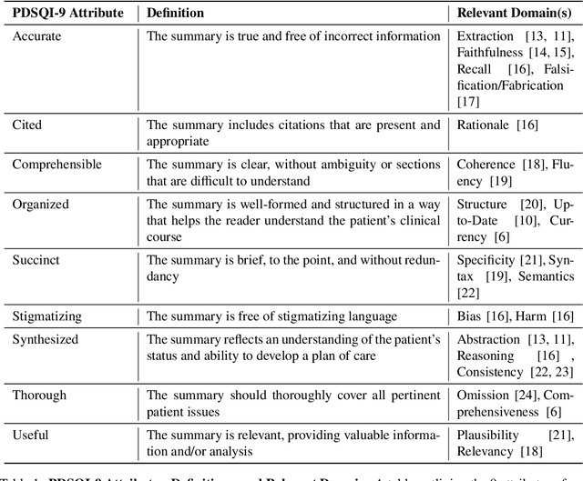
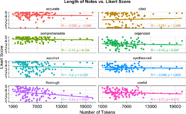

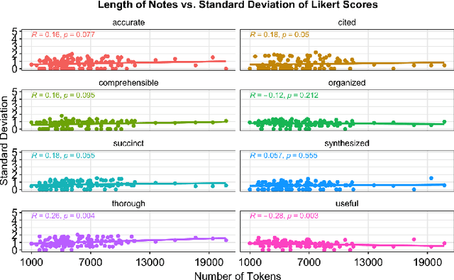
Abstract:As Large Language Models (LLMs) are integrated into electronic health record (EHR) workflows, validated instruments are essential to evaluate their performance before implementation. Existing instruments for provider documentation quality are often unsuitable for the complexities of LLM-generated text and lack validation on real-world data. The Provider Documentation Summarization Quality Instrument (PDSQI-9) was developed to evaluate LLM-generated clinical summaries. Multi-document summaries were generated from real-world EHR data across multiple specialties using several LLMs (GPT-4o, Mixtral 8x7b, and Llama 3-8b). Validation included Pearson correlation for substantive validity, factor analysis and Cronbach's alpha for structural validity, inter-rater reliability (ICC and Krippendorff's alpha) for generalizability, a semi-Delphi process for content validity, and comparisons of high- versus low-quality summaries for discriminant validity. Seven physician raters evaluated 779 summaries and answered 8,329 questions, achieving over 80% power for inter-rater reliability. The PDSQI-9 demonstrated strong internal consistency (Cronbach's alpha = 0.879; 95% CI: 0.867-0.891) and high inter-rater reliability (ICC = 0.867; 95% CI: 0.867-0.868), supporting structural validity and generalizability. Factor analysis identified a 4-factor model explaining 58% of the variance, representing organization, clarity, accuracy, and utility. Substantive validity was supported by correlations between note length and scores for Succinct (rho = -0.200, p = 0.029) and Organized (rho = -0.190, p = 0.037). Discriminant validity distinguished high- from low-quality summaries (p < 0.001). The PDSQI-9 demonstrates robust construct validity, supporting its use in clinical practice to evaluate LLM-generated summaries and facilitate safer integration of LLMs into healthcare workflows.
Particle-Filtering-based Latent Diffusion for Inverse Problems
Aug 25, 2024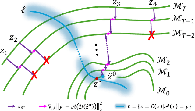

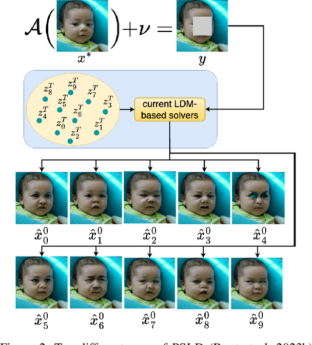

Abstract:Current strategies for solving image-based inverse problems apply latent diffusion models to perform posterior sampling.However, almost all approaches make no explicit attempt to explore the solution space, instead drawing only a single sample from a Gaussian distribution from which to generate their solution. In this paper, we introduce a particle-filtering-based framework for a nonlinear exploration of the solution space in the initial stages of reverse SDE methods. Our proposed particle-filtering-based latent diffusion (PFLD) method and proposed problem formulation and framework can be applied to any diffusion-based solution for linear or nonlinear inverse problems. Our experimental results show that PFLD outperforms the SoTA solver PSLD on the FFHQ-1K and ImageNet-1K datasets on inverse problem tasks of super resolution, Gaussian debluring and inpainting.
BIOSCAN-5M: A Multimodal Dataset for Insect Biodiversity
Jun 18, 2024


Abstract:As part of an ongoing worldwide effort to comprehend and monitor insect biodiversity, this paper presents the BIOSCAN-5M Insect dataset to the machine learning community and establish several benchmark tasks. BIOSCAN-5M is a comprehensive dataset containing multi-modal information for over 5 million insect specimens, and it significantly expands existing image-based biological datasets by including taxonomic labels, raw nucleotide barcode sequences, assigned barcode index numbers, and geographical information. We propose three benchmark experiments to demonstrate the impact of the multi-modal data types on the classification and clustering accuracy. First, we pretrain a masked language model on the DNA barcode sequences of the \mbox{BIOSCAN-5M} dataset, and demonstrate the impact of using this large reference library on species- and genus-level classification performance. Second, we propose a zero-shot transfer learning task applied to images and DNA barcodes to cluster feature embeddings obtained from self-supervised learning, to investigate whether meaningful clusters can be derived from these representation embeddings. Third, we benchmark multi-modality by performing contrastive learning on DNA barcodes, image data, and taxonomic information. This yields a general shared embedding space enabling taxonomic classification using multiple types of information and modalities. The code repository of the BIOSCAN-5M Insect dataset is available at {\url{https://github.com/zahrag/BIOSCAN-5M}}
A Step Towards Worldwide Biodiversity Assessment: The BIOSCAN-1M Insect Dataset
Jul 19, 2023Abstract:In an effort to catalog insect biodiversity, we propose a new large dataset of hand-labelled insect images, the BIOSCAN-Insect Dataset. Each record is taxonomically classified by an expert, and also has associated genetic information including raw nucleotide barcode sequences and assigned barcode index numbers, which are genetically-based proxies for species classification. This paper presents a curated million-image dataset, primarily to train computer-vision models capable of providing image-based taxonomic assessment, however, the dataset also presents compelling characteristics, the study of which would be of interest to the broader machine learning community. Driven by the biological nature inherent to the dataset, a characteristic long-tailed class-imbalance distribution is exhibited. Furthermore, taxonomic labelling is a hierarchical classification scheme, presenting a highly fine-grained classification problem at lower levels. Beyond spurring interest in biodiversity research within the machine learning community, progress on creating an image-based taxonomic classifier will also further the ultimate goal of all BIOSCAN research: to lay the foundation for a comprehensive survey of global biodiversity. This paper introduces the dataset and explores the classification task through the implementation and analysis of a baseline classifier.
Machine Learning Challenges of Biological Factors in Insect Image Data
Nov 04, 2022


Abstract:The BIOSCAN project, led by the International Barcode of Life Consortium, seeks to study changes in biodiversity on a global scale. One component of the project is focused on studying the species interaction and dynamics of all insects. In addition to genetically barcoding insects, over 1.5 million images per year will be collected, each needing taxonomic classification. With the immense volume of incoming images, relying solely on expert taxonomists to label the images would be impossible; however, artificial intelligence and computer vision technology may offer a viable high-throughput solution. Additional tasks including manually weighing individual insects to determine biomass, remain tedious and costly. Here again, computer vision may offer an efficient and compelling alternative. While the use of computer vision methods is appealing for addressing these problems, significant challenges resulting from biological factors present themselves. These challenges are formulated in the context of machine learning in this paper.
Normalized K-Means for Noise-Insensitive Multi-Dimensional Feature Learning
Feb 15, 2022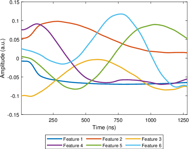
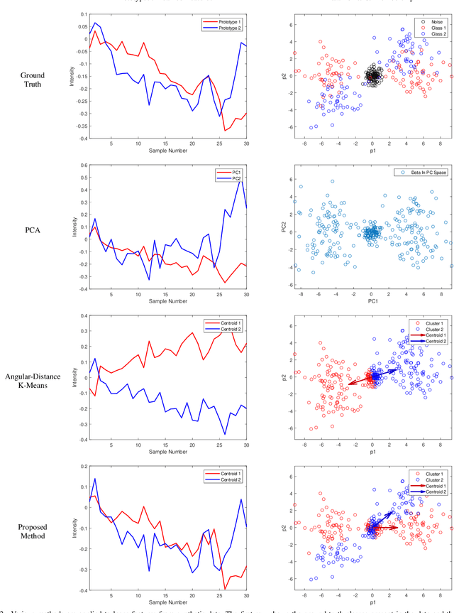
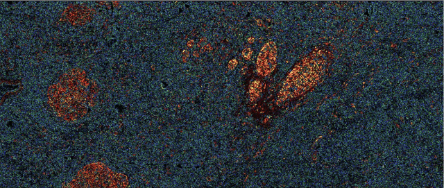
Abstract:Many measurement modalities which perform imaging by probing an object pixel-by-pixel, such as via Photoacoustic Microscopy, produce a multi-dimensional feature (typically a time-domain signal) at each pixel. In principle, the many degrees of freedom in the time-domain signal would admit the possibility of significant multi-modal information being implicitly present, much more than a single scalar "brightness", regarding the underlying targets being observed. However, the measured signal is neither a weighted-sum of basis functions (such as principal components) nor one of a set of prototypes (K-means), which has motivated the novel clustering method proposed here, capable of learning centroids (signal shapes) that are related to the underlying, albeit unknown, target characteristics in a scalable and noise-robust manner.
In vivo functional and structural retina imaging using multimodal photoacoustic remote sensing microscopy and optical coherence tomography
Aug 26, 2021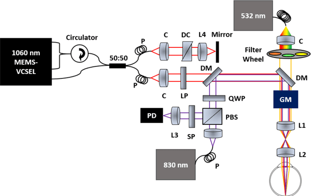
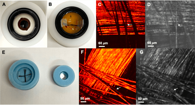
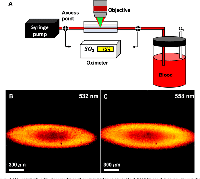
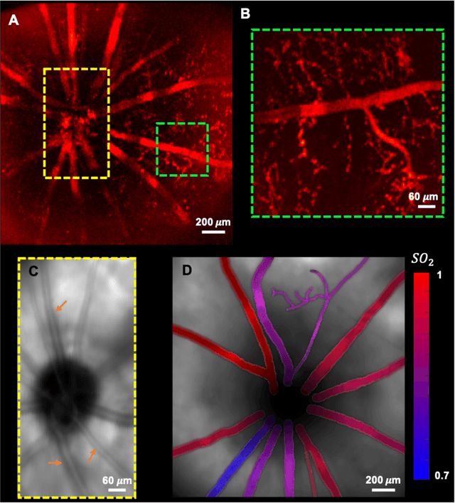
Abstract:We have developed a multimodal photoacoustic remote sensing (PARS) microscope combined with swept source optical coherence tomography for in vivo, non-contact retinal imaging. Building on the proven strength of multiwavelength PARS imaging, the system is applied for estimating retinal oxygen saturation in the rat retina. The capability of the technology is demonstrated by imaging both microanatomy and the microvasculature of the retina in vivo. To our knowledge this is the first time a non-contact photoacoustic imaging technique is employed for in vivo oxygen saturation measurement in the retina.
 Add to Chrome
 Add to Firefox
 Add to Edge