Nadieh Khalili
Navigating the landscape of multimodal AI in medicine: a scoping review on technical challenges and clinical applications
Nov 06, 2024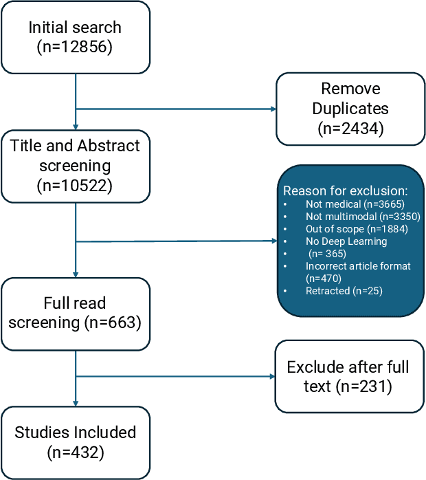
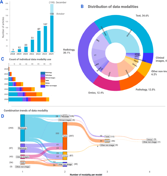
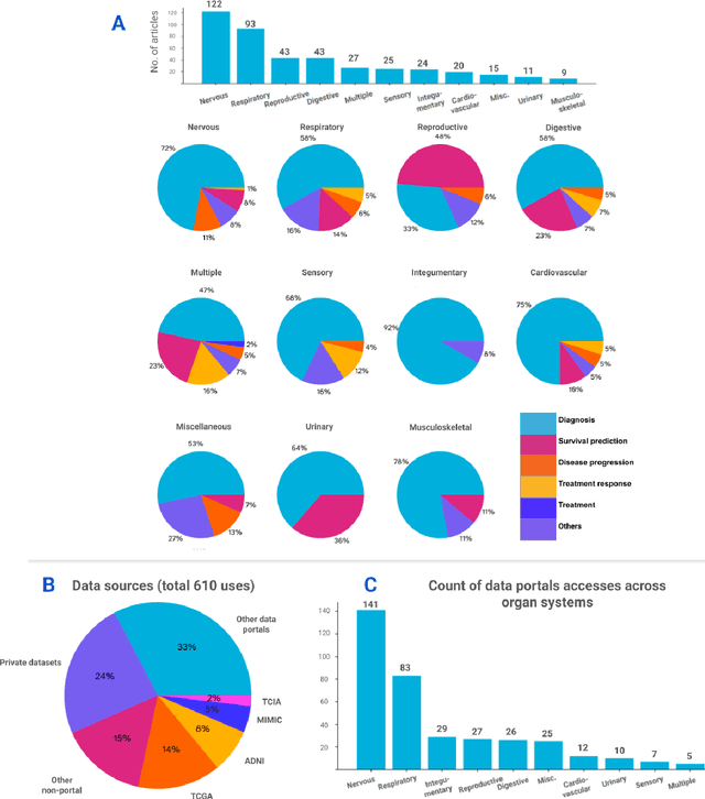
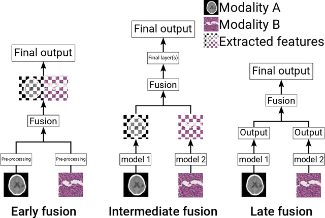
Abstract:Recent technological advances in healthcare have led to unprecedented growth in patient data quantity and diversity. While artificial intelligence (AI) models have shown promising results in analyzing individual data modalities, there is increasing recognition that models integrating multiple complementary data sources, so-called multimodal AI, could enhance clinical decision-making. This scoping review examines the landscape of deep learning-based multimodal AI applications across the medical domain, analyzing 432 papers published between 2018 and 2024. We provide an extensive overview of multimodal AI development across different medical disciplines, examining various architectural approaches, fusion strategies, and common application areas. Our analysis reveals that multimodal AI models consistently outperform their unimodal counterparts, with an average improvement of 6.2 percentage points in AUC. However, several challenges persist, including cross-departmental coordination, heterogeneous data characteristics, and incomplete datasets. We critically assess the technical and practical challenges in developing multimodal AI systems and discuss potential strategies for their clinical implementation, including a brief overview of commercially available multimodal AI models for clinical decision-making. Additionally, we identify key factors driving multimodal AI development and propose recommendations to accelerate the field's maturation. This review provides researchers and clinicians with a thorough understanding of the current state, challenges, and future directions of multimodal AI in medicine.
Structure-Preserving Multi-Domain Stain Color Augmentation using Style-Transfer with Disentangled Representations
Jul 26, 2021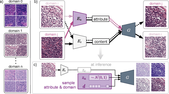

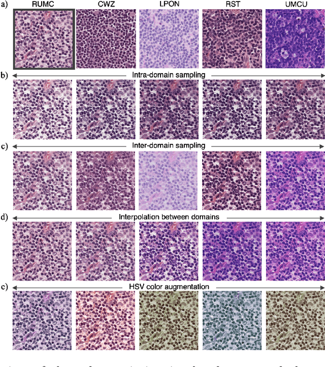
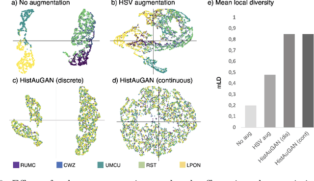
Abstract:In digital pathology, different staining procedures and scanners cause substantial color variations in whole-slide images (WSIs), especially across different laboratories. These color shifts result in a poor generalization of deep learning-based methods from the training domain to external pathology data. To increase test performance, stain normalization techniques are used to reduce the variance between training and test domain. Alternatively, color augmentation can be applied during training leading to a more robust model without the extra step of color normalization at test time. We propose a novel color augmentation technique, HistAuGAN, that can simulate a wide variety of realistic histology stain colors, thus making neural networks stain-invariant when applied during training. Based on a generative adversarial network (GAN) for image-to-image translation, our model disentangles the content of the image, i.e., the morphological tissue structure, from the stain color attributes. It can be trained on multiple domains and, therefore, learns to cover different stain colors as well as other domain-specific variations introduced in the slide preparation and imaging process. We demonstrate that HistAuGAN outperforms conventional color augmentation techniques on a classification task on the publicly available dataset Camelyon17 and show that it is able to mitigate present batch effects.
Combined analysis of coronary arteries and the left ventricular myocardium in cardiac CT angiography for detection of patients with functionally significant stenosis
Nov 10, 2019


Abstract:Treatment of patients with obstructive coronary artery disease is guided by the functional significance of a coronary artery stenosis. Fractional flow reserve (FFR), measured during invasive coronary angiography (ICA), is considered the gold standard to define the functional significance of a coronary stenosis. Here, we present a method for non-invasive detection of patients with functionally significant coronary artery stenosis, combining analysis of the coronary artery tree and the left ventricular (LV) myocardium in cardiac CT angiography (CCTA) images. We retrospectively collected CCTA scans of 126 patients who underwent invasive FFR measurements, to determine the functional significance of coronary stenoses. We combine our previous works for the analysis of the complete coronary artery tree and the LV myocardium: Coronary arteries are encoded by two disjoint convolutional autoencoders (CAEs) and the LV myocardium is characterized by a convolutional neural network (CNN) and a CAE. Thereafter, using the extracted encodings of all coronary arteries and the LV myocardium, patients are classified according to the presence of functionally significant stenosis, as defined by the invasively measured FFR. To handle the varying number of coronary arteries in a patient, the classification is formulated as a multiple instance learning problem and is performed using an attention-based neural network. Cross-validation experiments resulted in an average area under the receiver operating characteristic curve of $0.74 \pm 0.01$, and showed that the proposed combined analysis outperformed the analysis of the coronary arteries or the LV myocardium only. The results demonstrate the feasibility of combining the analyses of the complete coronary artery tree and the LV myocardium in CCTA images for the detection of patients with functionally significant stenosis in coronary arteries.
Deep learning analysis of cardiac CT angiography for detection of coronary arteries with functionally significant stenosis
Jun 11, 2019


Abstract:In patients with obstructive coronary artery disease, the functional significance of a coronary artery stenosis needs to be determined to guide treatment. This is typically established through fractional flow reserve (FFR) measurement, performed during invasive coronary angiography (ICA). We present a method for automatic and non-invasive detection of functionally significant coronary artery stenosis, employing deep unsupervised analysis of complete coronary arteries in cardiac CT angiography (CCTA) images. We retrospectively collected CCTA scans of 187 patients, 137 of them underwent invasive FFR measurement in 192 different coronary arteries. These FFR measurements served as a reference standard for the functional significance of the coronary stenosis. The centerlines of the coronary arteries were extracted and used to reconstruct straightened multi-planar reformatted (MPR) volumes. To automatically identify arteries with functionally significant stenosis, each MPR volume was encoded into a fixed number of encodings using two disjoint 3D and 1D convolutional autoencoders performing spatial and sequential encodings, respectively. Thereafter, these encodings were employed to classify arteries according to the presence of functionally significant stenosis using a support vector machine classifier. The detection of functionally significant stenosis, evaluated using repeated cross-validation experiments, resulted in an area under the receiver operating characteristic curve of $0.81 \pm 0.02$ on the artery-level, and $0.87 \pm 0.02$ on the patient-level. The results demonstrate that automatic non-invasive detection of the functionally significant stenosis in coronary arteries, using characteristics of complete coronary arteries in CCTA images, is feasible. This could potentially reduce the number of patients that unnecessarily undergo ICA.
 Add to Chrome
 Add to Firefox
 Add to Edge