Morteza Zabihi
Early Detection of Myocardial Infarction in Low-Quality Echocardiography
Oct 05, 2020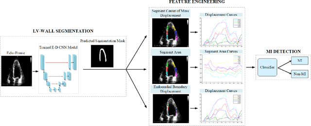
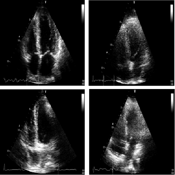

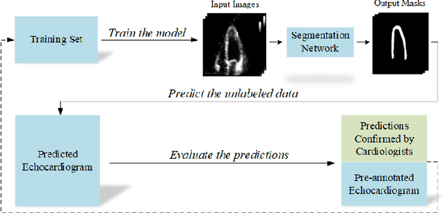
Abstract:Myocardial infarction (MI), or commonly known as heart attack, is a life-threatening worldwide health problem from which 32.4 million of people suffer each year. Early diagnosis and treatment of MI are crucial to prevent further heart tissue damages. However, MI detection in early stages is challenging because the symptoms are not easy to distinguish in electrocardiography findings or biochemical marker values found in the blood. Echocardiography is a noninvasive clinical tool for a more accurate early MI diagnosis, which is used to analyze the regional wall motion abnormalities. When echocardiography quality is poor, the diagnosis becomes a challenging and sometimes infeasible task even for a cardiologist. In this paper, we introduce a three-phase approach for early MI detection in low-quality echocardiography: 1) segmentation of the entire left ventricle (LV) wall of the heart using state-of-the-art deep learning model, 2) analysis of the segmented LV wall by feature engineering, and 3) early MI detection. The main contributions of this study are: highly accurate segmentation of the LV wall from low-resolution (both temporal and spatial) and noisy echocardiographic data, generating the segmentation ground-truth at pixel-level for the unannotated dataset using pseudo labeling approach, and composition of the first public echocardiographic dataset (HMC-QU) labeled by the cardiologists at the Hamad Medical Corporation Hospital in Qatar. Furthermore, the outputs of the proposed approach can significantly help cardiologists for a better assessment of the LV wall characteristics. The proposed method is evaluated in a 5-fold cross validation scheme on the HMC-QU dataset. The proposed approach has achieved an average level of 95.72% sensitivity and 99.58% specificity for the LV wall segmentation, and 85.97% sensitivity, 74.03% specificity, and 86.85% precision for MI detection.
Left Ventricular Wall Motion Estimation by Active Polynomials for Acute Myocardial Infarction Detection
Aug 11, 2020


Abstract:Echocardiogram (echo) is the earliest and the primary tool for identifying regional wall motion abnormalities (RWMA) in order to diagnose myocardial infarction (MI) or commonly known as heart attack. This paper proposes a novel approach, Active Polynomials, which can accurately and robustly estimate the global motion of the Left Ventricular (LV) wall from any echo in a robust and accurate way. The proposed algorithm quantifies the true wall motion occurring in LV wall segments so as to assist cardiologists diagnose early signs of an acute MI. It further enables medical experts to gain an enhanced visualization capability of echo images through color-coded segments along with their "maximum motion displacement" plots helping them to better assess wall motion and LV Ejection-Fraction (LVEF). The outputs of the method can further help echo-technicians to assess and improve the quality of the echocardiogram recording. A major contribution of this study is the first public echo database collection composed by physicians at the Hamad Medical Corporation Hospital in Qatar. The so-called HMC-QU database will serve as the benchmark for the forthcoming relevant studies. The results over the HMC-QU dataset show that the proposed approach can achieve high accuracy, sensitivity and precision in MI detection even though the echo quality is quite poor, and the temporal resolution is low.
Automated Polysomnography Analysis for Detection of Non-Apneic and Non-Hypopneic Arousals using Feature Engineering and a Bidirectional LSTM Network
Sep 06, 2019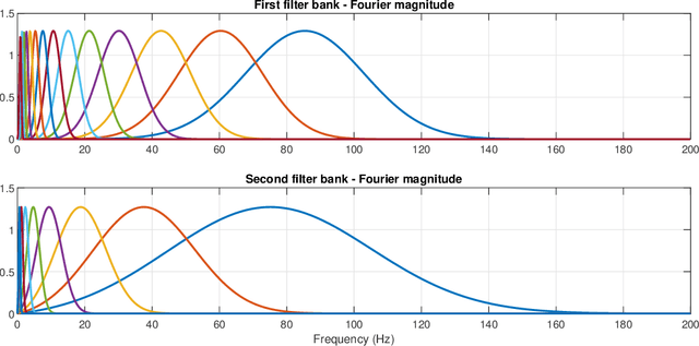

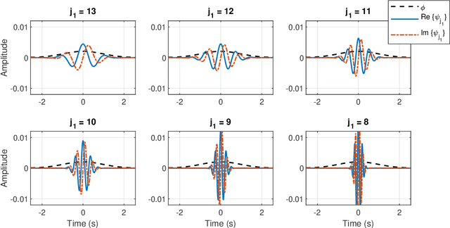
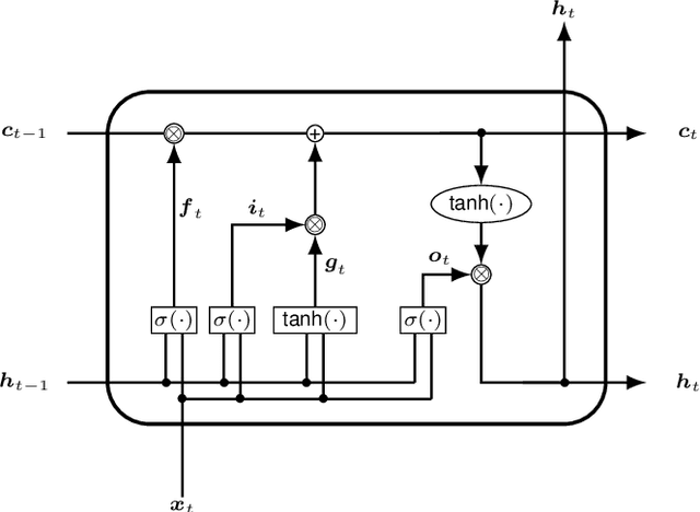
Abstract:Objective: The aim of this study is to develop an automated classification algorithm for polysomnography (PSG) recordings to detect non-apneic and non-hypopneic arousals. Our particular focus is on detecting the respiratory effort-related arousals (RERAs) which are very subtle respiratory events that do not meet the criteria for apnea or hypopnea, and are more challenging to detect. Methods: The proposed algorithm is based on a bidirectional long short-term memory (BiLSTM) classifier and 465 multi-domain features, extracted from multimodal clinical time series. The features consist of a set of physiology-inspired features (n = 75), obtained by multiple steps of feature selection and expert analysis, and a set of physiology-agnostic features (n = 390), derived from scattering transform. Results: The proposed algorithm is validated on the 2018 PhysioNet challenge dataset. The overall performance in terms of the area under the precision-recall curve (AUPRC) is 0.50 on the hidden test dataset. This result is tied for the second-best score during the follow-up and official phases of the 2018 PhysioNet challenge. Conclusions: The results demonstrate that it is possible to automatically detect subtle non-apneic/non-hypopneic arousal events from PSG recordings. Significance: Automatic detection of subtle respiratory events such as RERAs together with other non-apneic/non-hypopneic arousals will allow detailed annotations of large PSG databases. This contributes to a better retrospective analysis of sleep data, which may also improve the quality of treatment.
1D Convolutional Neural Network Models for Sleep Arousal Detection
Mar 01, 2019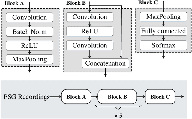
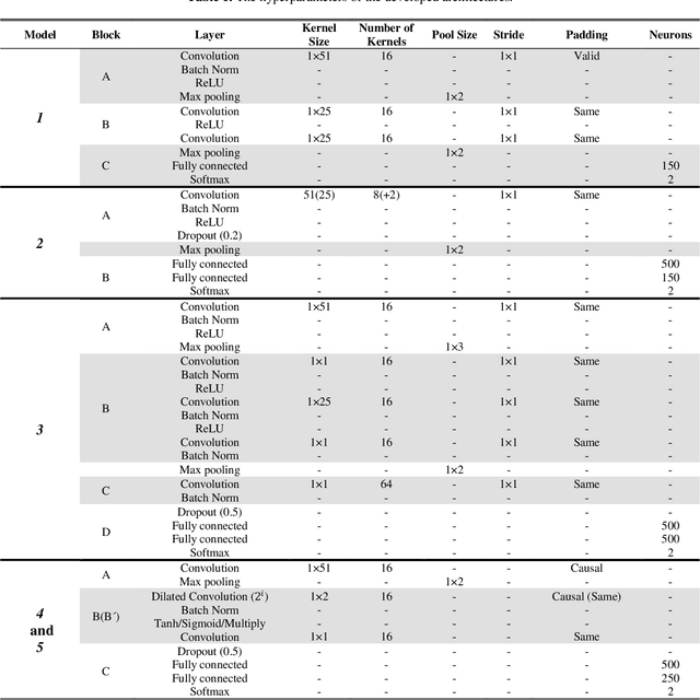
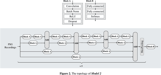
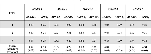
Abstract:Sleep arousals transition the depth of sleep to a more superficial stage. The occurrence of such events is often considered as a protective mechanism to alert the body of harmful stimuli. Thus, accurate sleep arousal detection can lead to an enhanced understanding of the underlying causes and influencing the assessment of sleep quality. Previous studies and guidelines have suggested that sleep arousals are linked mainly to abrupt frequency shifts in EEG signals, but the proposed rules are shown to be insufficient for a comprehensive characterization of arousals. This study investigates the application of five recent convolutional neural networks (CNNs) for sleep arousal detection and performs comparative evaluations to determine the best model for this task. The investigated state-of-the-art CNN models have originally been designed for image or speech processing. A detailed set of evaluations is performed on the benchmark dataset provided by PhysioNet/Computing in Cardiology Challenge 2018, and the results show that the best 1D CNN model has achieved an average of 0.31 and 0.84 for the area under the precision-recall and area under the ROC curves, respectively.
 Add to Chrome
 Add to Firefox
 Add to Edge