Matteo Figini
Centre for Medical Image Computing and Department of Computer Science - University College London - UK
Tackling Hallucination from Conditional Models for Medical Image Reconstruction with DynamicDPS
Mar 03, 2025Abstract:Hallucinations are spurious structures not present in the ground truth, posing a critical challenge in medical image reconstruction, especially for data-driven conditional models. We hypothesize that combining an unconditional diffusion model with data consistency, trained on a diverse dataset, can reduce these hallucinations. Based on this, we propose DynamicDPS, a diffusion-based framework that integrates conditional and unconditional diffusion models to enhance low-quality medical images while systematically reducing hallucinations. Our approach first generates an initial reconstruction using a conditional model, then refines it with an adaptive diffusion-based inverse problem solver. DynamicDPS skips early stage in the reverse process by selecting an optimal starting time point per sample and applies Wolfe's line search for adaptive step sizes, improving both efficiency and image fidelity. Using diffusion priors and data consistency, our method effectively reduces hallucinations from any conditional model output. We validate its effectiveness in Image Quality Transfer for low-field MRI enhancement. Extensive evaluations on synthetic and real MR scans, including a downstream task for tissue volume estimation, show that DynamicDPS reduces hallucinations, improving relative volume estimation by over 15% for critical tissues while using only 5% of the sampling steps required by baseline diffusion models. As a model-agnostic and fine-tuning-free approach, DynamicDPS offers a robust solution for hallucination reduction in medical imaging. The code will be made publicly available upon publication.
Alternative Learning Paradigms for Image Quality Transfer
Nov 08, 2024


Abstract:Image Quality Transfer (IQT) aims to enhance the contrast and resolution of low-quality medical images, e.g. obtained from low-power devices, with rich information learned from higher quality images. In contrast to existing IQT methods which adopt supervised learning frameworks, in this work, we propose two novel formulations of the IQT problem. The first approach uses an unsupervised learning framework, whereas the second is a combination of both supervised and unsupervised learning. The unsupervised learning approach considers a sparse representation (SRep) and dictionary learning model, which we call IQT-SRep, whereas the combination of supervised and unsupervised learning approach is based on deep dictionary learning (DDL), which we call IQT-DDL. The IQT-SRep approach trains two dictionaries using a SRep model using pairs of low- and high-quality volumes. Subsequently, the SRep of a low-quality block, in terms of the low-quality dictionary, can be directly used to recover the corresponding high-quality block using the high-quality dictionary. On the other hand, the IQT-DDL approach explicitly learns a high-resolution dictionary to upscale the input volume, while the entire network, including high dictionary generator, is simultaneously optimised to take full advantage of deep learning methods. The two models are evaluated using a low-field magnetic resonance imaging (MRI) application aiming to recover high-quality images akin to those obtained from high-field scanners. Experiments comparing the proposed approaches against state-of-the-art supervised deep learning IQT method (IQT-DL) identify that the two novel formulations of the IQT problem can avoid bias associated with supervised methods when tested using out-of-distribution data that differs from the distribution of the data the model was trained on. This highlights the potential benefit of these novel paradigms for IQT.
* Accepted for publication at the Journal of Machine Learning for Biomedical Imaging (MELBA) https://melba-journal.org/2024:027
Image Quality Transfer of Diffusion MRI Guided By High-Resolution Structural MRI
Aug 06, 2024


Abstract:Prior work on the Image Quality Transfer on Diffusion MRI (dMRI) has shown significant improvement over traditional interpolation methods. However, the difficulty in obtaining ultra-high resolution Diffusion MRI scans poses a problem in training neural networks to obtain high-resolution dMRI scans. Here we hypothesise that the inclusion of structural MRI images, which can be acquired at much higher resolutions, can be used as a guide to obtaining a more accurate high-resolution dMRI output. To test our hypothesis, we have constructed a novel framework that incorporates structural MRI scans together with dMRI to obtain high-resolution dMRI scans. We set up tests which evaluate the validity of our claim through various configurations and compare the performance of our approach against a unimodal approach. Our results show that the inclusion of structural MRI scans do lead to an improvement in high-resolution image prediction when T1w data is incorporated into the model input.
Tackling Structural Hallucination in Image Translation with Local Diffusion
Apr 13, 2024


Abstract:Recent developments in diffusion models have advanced conditioned image generation, yet they struggle with reconstructing out-of-distribution (OOD) images, such as unseen tumors in medical images, causing ``image hallucination'' and risking misdiagnosis. We hypothesize such hallucinations result from local OOD regions in the conditional images. We verify that partitioning the OOD region and conducting separate image generations alleviates hallucinations in several applications. From this, we propose a training-free diffusion framework that reduces hallucination with multiple Local Diffusion processes. Our approach involves OOD estimation followed by two modules: a ``branching'' module generates locally both within and outside OOD regions, and a ``fusion'' module integrates these predictions into one. Our evaluation shows our method mitigates hallucination over baseline models quantitatively and qualitatively, reducing misdiagnosis by 40% and 25% in the real-world medical and natural image datasets, respectively. It also demonstrates compatibility with various pre-trained diffusion models.
A 3D Conditional Diffusion Model for Image Quality Transfer -- An Application to Low-Field MRI
Nov 11, 2023


Abstract:Low-field (LF) MRI scanners (<1T) are still prevalent in settings with limited resources or unreliable power supply. However, they often yield images with lower spatial resolution and contrast than high-field (HF) scanners. This quality disparity can result in inaccurate clinician interpretations. Image Quality Transfer (IQT) has been developed to enhance the quality of images by learning a mapping function between low and high-quality images. Existing IQT models often fail to restore high-frequency features, leading to blurry output. In this paper, we propose a 3D conditional diffusion model to improve 3D volumetric data, specifically LF MR images. Additionally, we incorporate a cross-batch mechanism into the self-attention and padding of our network, ensuring broader contextual awareness even under small 3D patches. Experiments on the publicly available Human Connectome Project (HCP) dataset for IQT and brain parcellation demonstrate that our model outperforms existing methods both quantitatively and qualitatively. The code is publicly available at \url{https://github.com/edshkim98/DiffusionIQT}.
Low-field magnetic resonance image enhancement via stochastic image quality transfer
Apr 26, 2023


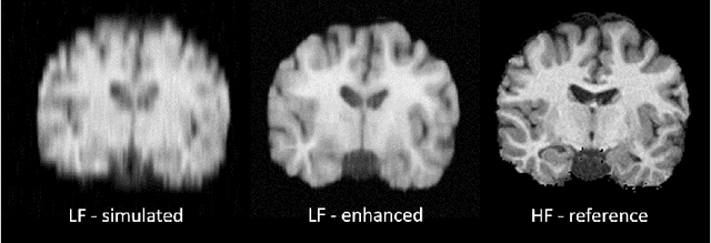
Abstract:Low-field (<1T) magnetic resonance imaging (MRI) scanners remain in widespread use in low- and middle-income countries (LMICs) and are commonly used for some applications in higher income countries e.g. for small child patients with obesity, claustrophobia, implants, or tattoos. However, low-field MR images commonly have lower resolution and poorer contrast than images from high field (1.5T, 3T, and above). Here, we present Image Quality Transfer (IQT) to enhance low-field structural MRI by estimating from a low-field image the image we would have obtained from the same subject at high field. Our approach uses (i) a stochastic low-field image simulator as the forward model to capture uncertainty and variation in the contrast of low-field images corresponding to a particular high-field image, and (ii) an anisotropic U-Net variant specifically designed for the IQT inverse problem. We evaluate the proposed algorithm both in simulation and using multi-contrast (T1-weighted, T2-weighted, and fluid attenuated inversion recovery (FLAIR)) clinical low-field MRI data from an LMIC hospital. We show the efficacy of IQT in improving contrast and resolution of low-field MR images. We demonstrate that IQT-enhanced images have potential for enhancing visualisation of anatomical structures and pathological lesions of clinical relevance from the perspective of radiologists. IQT is proved to have capability of boosting the diagnostic value of low-field MRI, especially in low-resource settings.
Deformably-Scaled Transposed Convolution
Oct 17, 2022


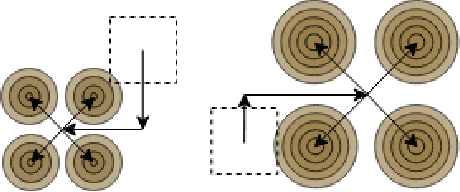
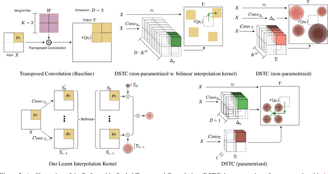
Abstract:Transposed convolution is crucial for generating high-resolution outputs, yet has received little attention compared to convolution layers. In this work we revisit transposed convolution and introduce a novel layer that allows us to place information in the image selectively and choose the `stroke breadth' at which the image is synthesized, whilst incurring a small additional parameter cost. For this we introduce three ideas: firstly, we regress offsets to the positions where the transpose convolution results are placed; secondly we broadcast the offset weight locations over a learnable neighborhood; and thirdly we use a compact parametrization to share weights and restrict offsets. We show that simply substituting upsampling operators with our novel layer produces substantial improvements across tasks as diverse as instance segmentation, object detection, semantic segmentation, generative image modeling, and 3D magnetic resonance image enhancement, while outperforming all existing variants of transposed convolutions. Our novel layer can be used as a drop-in replacement for 2D and 3D upsampling operators and the code will be publicly available.
Progressive Subsampling for Oversampled Data -- Application to Quantitative MRI
Apr 08, 2022


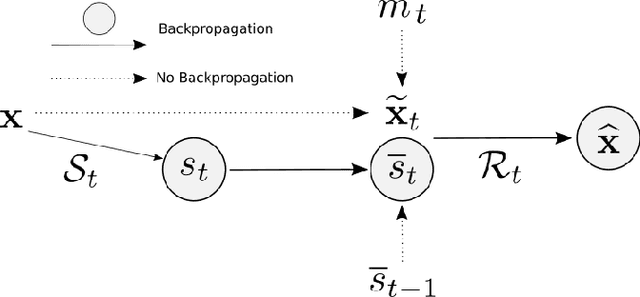
Abstract:We present PROSUB: PROgressive SUBsampling, a deep learning based, automated methodology that subsamples an oversampled data set (e.g. multi-channeled 3D images) with minimal loss of information. We build upon a recent dual-network approach that won the MICCAI MUlti-DIffusion (MUDI) quantitative MRI measurement sampling-reconstruction challenge, but suffers from deep learning training instability, by subsampling with a hard decision boundary. PROSUB uses the paradigm of recursive feature elimination (RFE) and progressively subsamples measurements during deep learning training, improving optimization stability. PROSUB also integrates a neural architecture search (NAS) paradigm, allowing the network architecture hyperparameters to respond to the subsampling process. We show PROSUB outperforms the winner of the MUDI MICCAI challenge, producing large improvements >18% MSE on the MUDI challenge sub-tasks and qualitative improvements on downstream processes useful for clinical applications. We also show the benefits of incorporating NAS and analyze the effect of PROSUB's components. As our method generalizes to other problems beyond MRI measurement selection-reconstruction, our code is https://github.com/sbb-gh/PROSUB
Image Quality Transfer Enhances Contrast and Resolution of Low-Field Brain MRI in African Paediatric Epilepsy Patients
Mar 18, 2020


Abstract:1.5T or 3T scanners are the current standard for clinical MRI, but low-field (<1T) scanners are still common in many lower- and middle-income countries for reasons of cost and robustness to power failures. Compared to modern high-field scanners, low-field scanners provide images with lower signal-to-noise ratio at equivalent resolution, leaving practitioners to compensate by using large slice thickness and incomplete spatial coverage. Furthermore, the contrast between different types of brain tissue may be substantially reduced even at equal signal-to-noise ratio, which limits diagnostic value. Recently the paradigm of Image Quality Transfer has been applied to enhance 0.36T structural images aiming to approximate the resolution, spatial coverage, and contrast of typical 1.5T or 3T images. A variant of the neural network U-Net was trained using low-field images simulated from the publicly available 3T Human Connectome Project dataset. Here we present qualitative results from real and simulated clinical low-field brain images showing the potential value of IQT to enhance the clinical utility of readily accessible low-field MRIs in the management of epilepsy.
Deep Learning for Low-Field to High-Field MR: Image Quality Transfer with Probabilistic Decimation Simulator
Sep 15, 2019


Abstract:MR images scanned at low magnetic field ($<1$T) have lower resolution in the slice direction and lower contrast, due to a relatively small signal-to-noise ratio (SNR) than those from high field (typically 1.5T and 3T). We adapt the recent idea of Image Quality Transfer (IQT) to enhance very low-field structural images aiming to estimate the resolution, spatial coverage, and contrast of high-field images. Analogous to many learning-based image enhancement techniques, IQT generates training data from high-field scans alone by simulating low-field images through a pre-defined decimation model. However, the ground truth decimation model is not well-known in practice, and lack of its specification can bias the trained model, aggravating performance on the real low-field scans. In this paper we propose a probabilistic decimation simulator to improve robustness of model training. It is used to generate and augment various low-field images whose parameters are random variables and sampled from an empirical distribution related to tissue-specific SNR on a 0.36T scanner. The probabilistic decimation simulator is model-agnostic, that is, it can be used with any super-resolution networks. Furthermore we propose a variant of U-Net architecture to improve its learning performance. We show promising qualitative results from clinical low-field images confirming the strong efficacy of IQT in an important new application area: epilepsy diagnosis in sub-Saharan Africa where only low-field scanners are normally available.
 Add to Chrome
 Add to Firefox
 Add to Edge