Lijun Lu
Unveiling and Bridging the Functional Perception Gap in MLLMs: Atomic Visual Alignment and Hierarchical Evaluation via PET-Bench
Jan 06, 2026Abstract:While Multimodal Large Language Models (MLLMs) have demonstrated remarkable proficiency in tasks such as abnormality detection and report generation for anatomical modalities, their capability in functional imaging remains largely unexplored. In this work, we identify and quantify a fundamental functional perception gap: the inability of current vision encoders to decode functional tracer biodistribution independent of morphological priors. Identifying Positron Emission Tomography (PET) as the quintessential modality to investigate this disconnect, we introduce PET-Bench, the first large-scale functional imaging benchmark comprising 52,308 hierarchical QA pairs from 9,732 multi-site, multi-tracer PET studies. Extensive evaluation of 19 state-of-the-art MLLMs reveals a critical safety hazard termed the Chain-of-Thought (CoT) hallucination trap. We observe that standard CoT prompting, widely considered to enhance reasoning, paradoxically decouples linguistic generation from visual evidence in PET, producing clinically fluent but factually ungrounded diagnoses. To resolve this, we propose Atomic Visual Alignment (AVA), a simple fine-tuning strategy that enforces the mastery of low-level functional perception prior to high-level diagnostic reasoning. Our results demonstrate that AVA effectively bridges the perception gap, transforming CoT from a source of hallucination into a robust inference tool and improving diagnostic accuracy by up to 14.83%. Code and data are available at https://github.com/yezanting/PET-Bench.
Kimi K2: Open Agentic Intelligence
Jul 28, 2025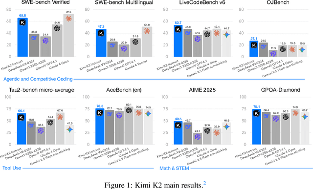


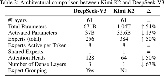
Abstract:We introduce Kimi K2, a Mixture-of-Experts (MoE) large language model with 32 billion activated parameters and 1 trillion total parameters. We propose the MuonClip optimizer, which improves upon Muon with a novel QK-clip technique to address training instability while enjoying the advanced token efficiency of Muon. Based on MuonClip, K2 was pre-trained on 15.5 trillion tokens with zero loss spike. During post-training, K2 undergoes a multi-stage post-training process, highlighted by a large-scale agentic data synthesis pipeline and a joint reinforcement learning (RL) stage, where the model improves its capabilities through interactions with real and synthetic environments. Kimi K2 achieves state-of-the-art performance among open-source non-thinking models, with strengths in agentic capabilities. Notably, K2 obtains 66.1 on Tau2-Bench, 76.5 on ACEBench (En), 65.8 on SWE-Bench Verified, and 47.3 on SWE-Bench Multilingual -- surpassing most open and closed-sourced baselines in non-thinking settings. It also exhibits strong capabilities in coding, mathematics, and reasoning tasks, with a score of 53.7 on LiveCodeBench v6, 49.5 on AIME 2025, 75.1 on GPQA-Diamond, and 27.1 on OJBench, all without extended thinking. These results position Kimi K2 as one of the most capable open-source large language models to date, particularly in software engineering and agentic tasks. We release our base and post-trained model checkpoints to facilitate future research and applications of agentic intelligence.
MDAA-Diff: CT-Guided Multi-Dose Adaptive Attention Diffusion Model for PET Denoising
May 08, 2025Abstract:Acquiring high-quality Positron Emission Tomography (PET) images requires administering high-dose radiotracers, which increases radiation exposure risks. Generating standard-dose PET (SPET) from low-dose PET (LPET) has become a potential solution. However, previous studies have primarily focused on single low-dose PET denoising, neglecting two critical factors: discrepancies in dose response caused by inter-patient variability, and complementary anatomical constraints derived from CT images. In this work, we propose a novel CT-Guided Multi-dose Adaptive Attention Denoising Diffusion Model (MDAA-Diff) for multi-dose PET denoising. Our approach integrates anatomical guidance and dose-level adaptation to achieve superior denoising performance under low-dose conditions. Specifically, this approach incorporates a CT-Guided High-frequency Wavelet Attention (HWA) module, which uses wavelet transforms to separate high-frequency anatomical boundary features from CT images. These extracted features are then incorporated into PET imaging through an adaptive weighted fusion mechanism to enhance edge details. Additionally, we propose the Dose-Adaptive Attention (DAA) module, a dose-conditioned enhancement mechanism that dynamically integrates dose levels into channel-spatial attention weight calculation. Extensive experiments on 18F-FDG and 68Ga-FAPI datasets demonstrate that MDAA-Diff outperforms state-of-the-art approaches in preserving diagnostic quality under reduced-dose conditions. Our code is publicly available.
Semi-KAN: KAN Provides an Effective Representation for Semi-Supervised Learning in Medical Image Segmentation
Mar 19, 2025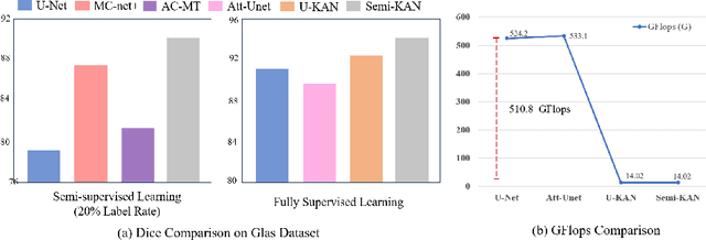
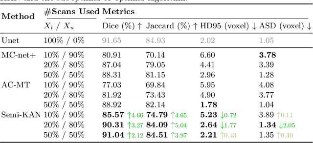
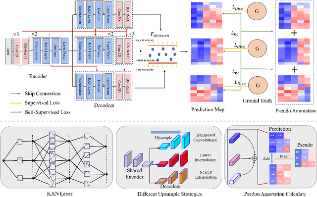
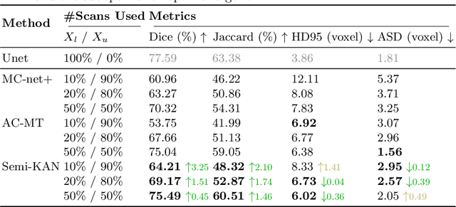
Abstract:Deep learning-based medical image segmentation has shown remarkable success; however, it typically requires extensive pixel-level annotations, which are both expensive and time-intensive. Semi-supervised medical image segmentation (SSMIS) offers a viable alternative, driven by advancements in CNNs and ViTs. However, these networks often rely on single fixed activation functions and linear modeling patterns, limiting their ability to effectively learn robust representations. Given the limited availability of labeled date, achieving robust representation learning becomes crucial. Inspired by Kolmogorov-Arnold Networks (KANs), we propose Semi-KAN, which leverages the untapped potential of KANs to enhance backbone architectures for representation learning in SSMIS. Our findings indicate that: (1) compared to networks with fixed activation functions, KANs exhibit superior representation learning capabilities with fewer parameters, and (2) KANs excel in high-semantic feature spaces. Building on these insights, we integrate KANs into tokenized intermediate representations, applying them selectively at the encoder's bottleneck and the decoder's top layers within a U-Net pipeline to extract high-level semantic features. Although learnable activation functions improve feature expansion, they introduce significant computational overhead with only marginal performance gains. To mitigate this, we reduce the feature dimensions and employ horizontal scaling to capture multiple pattern representations. Furthermore, we design a multi-branch U-Net architecture with uncertainty estimation to effectively learn diverse pattern representations. Extensive experiments on four public datasets demonstrate that Semi-KAN surpasses baseline networks, utilizing fewer KAN layers and lower computational cost, thereby underscoring the potential of KANs as a promising approach for SSMIS.
Self is the Best Learner: CT-free Ultra-Low-Dose PET Organ Segmentation via Collaborating Denoising and Segmentation Learning
Mar 05, 2025Abstract:Organ segmentation in Positron Emission Tomography (PET) plays a vital role in cancer quantification. Low-dose PET (LDPET) provides a safer alternative by reducing radiation exposure. However, the inherent noise and blurred boundaries make organ segmentation more challenging. Additionally, existing PET organ segmentation methods rely on co-registered Computed Tomography (CT) annotations, overlooking the problem of modality mismatch. In this study, we propose LDOS, a novel CT-free ultra-LDPET organ segmentation pipeline. Inspired by Masked Autoencoders (MAE), we reinterpret LDPET as a naturally masked version of Full-Dose PET (FDPET). LDOS adopts a simple yet effective architecture: a shared encoder extracts generalized features, while task-specific decoders independently refine outputs for denoising and segmentation. By integrating CT-derived organ annotations into the denoising process, LDOS improves anatomical boundary recognition and alleviates the PET/CT misalignments. Experiments demonstrate that LDOS achieves state-of-the-art performance with mean Dice scores of 73.11% (18F-FDG) and 73.97% (68Ga-FAPI) across 18 organs in 5% dose PET. Our code is publicly available.
A Multi-Scale Spatial Transformer U-Net for Simultaneously Automatic Reorientation and Segmentation of 3D Nuclear Cardiac Images
Oct 16, 2023Abstract:Accurate reorientation and segmentation of the left ventricular (LV) is essential for the quantitative analysis of myocardial perfusion imaging (MPI), in which one critical step is to reorient the reconstructed transaxial nuclear cardiac images into standard short-axis slices for subsequent image processing. Small-scale LV myocardium (LV-MY) region detection and the diverse cardiac structures of individual patients pose challenges to LV segmentation operation. To mitigate these issues, we propose an end-to-end model, named as multi-scale spatial transformer UNet (MS-ST-UNet), that involves the multi-scale spatial transformer network (MSSTN) and multi-scale UNet (MSUNet) modules to perform simultaneous reorientation and segmentation of LV region from nuclear cardiac images. The proposed method is trained and tested using two different nuclear cardiac image modalities: 13N-ammonia PET and 99mTc-sestamibi SPECT. We use a multi-scale strategy to generate and extract image features with different scales. Our experimental results demonstrate that the proposed method significantly improves the reorientation and segmentation performance. This joint learning framework promotes mutual enhancement between reorientation and segmentation tasks, leading to cutting edge performance and an efficient image processing workflow. The proposed end-to-end deep network has the potential to reduce the burden of manual delineation for cardiac images, thereby providing multimodal quantitative analysis assistance for physicists.
Joint nnU-Net and Radiomics Approaches for Segmentation and Prognosis of Head and Neck Cancers with PET/CT images
Nov 18, 2022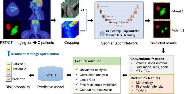
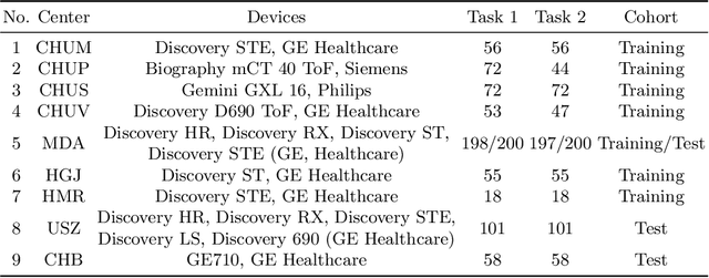
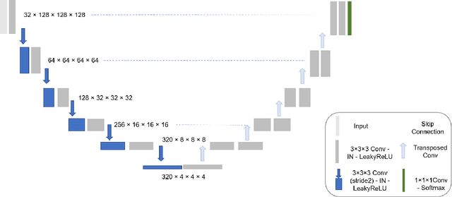

Abstract:Automatic segmentation of head and neck cancer (HNC) tumors and lymph nodes plays a crucial role in the optimization treatment strategy and prognosis analysis. This study aims to employ nnU-Net for automatic segmentation and radiomics for recurrence-free survival (RFS) prediction using pretreatment PET/CT images in multi-center HNC cohort. A multi-center HNC dataset with 883 patients (524 patients for training, 359 for testing) was provided in HECKTOR 2022. A bounding box of the extended oropharyngeal region was retrieved for each patient with fixed size of 224 x 224 x 224 $mm^{3}$. Then 3D nnU-Net architecture was adopted to automatic segmentation of primary tumor and lymph nodes synchronously.Based on predicted segmentation, ten conventional features and 346 standardized radiomics features were extracted for each patient. Three prognostic models were constructed containing conventional and radiomics features alone, and their combinations by multivariate CoxPH modelling. The statistical harmonization method, ComBat, was explored towards reducing multicenter variations. Dice score and C-index were used as evaluation metrics for segmentation and prognosis task, respectively. For segmentation task, we achieved mean dice score around 0.701 for primary tumor and lymph nodes by 3D nnU-Net. For prognostic task, conventional and radiomics models obtained the C-index of 0.658 and 0.645 in the test set, respectively, while the combined model did not improve the prognostic performance with the C-index of 0.648.
 Add to Chrome
 Add to Firefox
 Add to Edge