Ellen M. Mowry
MSRepaint: Multiple Sclerosis Repaint with Conditional Denoising Diffusion Implicit Model for Bidirectional Lesion Filling and Synthesis
Oct 02, 2025Abstract:In multiple sclerosis, lesions interfere with automated magnetic resonance imaging analyses such as brain parcellation and deformable registration, while lesion segmentation models are hindered by the limited availability of annotated training data. To address both issues, we propose MSRepaint, a unified diffusion-based generative model for bidirectional lesion filling and synthesis that restores anatomical continuity for downstream analyses and augments segmentation through realistic data generation. MSRepaint conditions on spatial lesion masks for voxel-level control, incorporates contrast dropout to handle missing inputs, integrates a repainting mechanism to preserve surrounding anatomy during lesion filling and synthesis, and employs a multi-view DDIM inversion and fusion pipeline for 3D consistency with fast inference. Extensive evaluations demonstrate the effectiveness of MSRepaint across multiple tasks. For lesion filling, we evaluate both the accuracy within the filled regions and the impact on downstream tasks including brain parcellation and deformable registration. MSRepaint outperforms the traditional lesion filling methods FSL and NiftySeg, and achieves accuracy on par with FastSurfer-LIT, a recent diffusion model-based inpainting method, while offering over 20 times faster inference. For lesion synthesis, state-of-the-art MS lesion segmentation models trained on MSRepaint-synthesized data outperform those trained on CarveMix-synthesized data or real ISBI challenge training data across multiple benchmarks, including the MICCAI 2016 and UMCL datasets. Additionally, we demonstrate that MSRepaint's unified bidirectional filling and synthesis capability, with full spatial control over lesion appearance, enables high-fidelity simulation of lesion evolution in longitudinal MS progression.
UNISELF: A Unified Network with Instance Normalization and Self-Ensembled Lesion Fusion for Multiple Sclerosis Lesion Segmentation
Aug 06, 2025
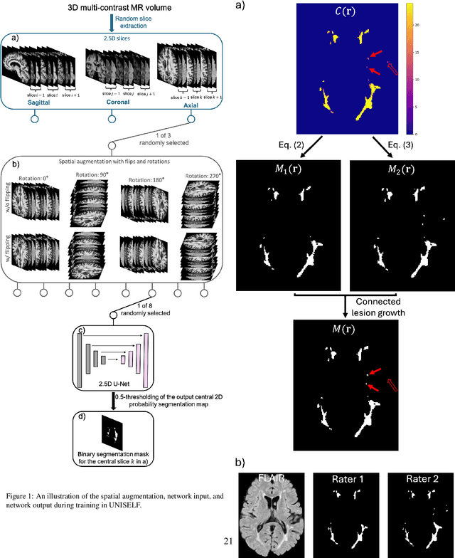
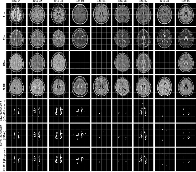

Abstract:Automated segmentation of multiple sclerosis (MS) lesions using multicontrast magnetic resonance (MR) images improves efficiency and reproducibility compared to manual delineation, with deep learning (DL) methods achieving state-of-the-art performance. However, these DL-based methods have yet to simultaneously optimize in-domain accuracy and out-of-domain generalization when trained on a single source with limited data, or their performance has been unsatisfactory. To fill this gap, we propose a method called UNISELF, which achieves high accuracy within a single training domain while demonstrating strong generalizability across multiple out-of-domain test datasets. UNISELF employs a novel test-time self-ensembled lesion fusion to improve segmentation accuracy, and leverages test-time instance normalization (TTIN) of latent features to address domain shifts and missing input contrasts. Trained on the ISBI 2015 longitudinal MS segmentation challenge training dataset, UNISELF ranks among the best-performing methods on the challenge test dataset. Additionally, UNISELF outperforms all benchmark methods trained on the same ISBI training data across diverse out-of-domain test datasets with domain shifts and missing contrasts, including the public MICCAI 2016 and UMCL datasets, as well as a private multisite dataset. These test datasets exhibit domain shifts and/or missing contrasts caused by variations in acquisition protocols, scanner types, and imaging artifacts arising from imperfect acquisition. Our code is available at https://github.com/uponacceptance.
Synthetic multi-inversion time magnetic resonance images for visualization of subcortical structures
Jun 04, 2025Abstract:Purpose: Visualization of subcortical gray matter is essential in neuroscience and clinical practice, particularly for disease understanding and surgical planning.While multi-inversion time (multi-TI) T$_1$-weighted (T$_1$-w) magnetic resonance (MR) imaging improves visualization, it is rarely acquired in clinical settings. Approach: We present SyMTIC (Synthetic Multi-TI Contrasts), a deep learning method that generates synthetic multi-TI images using routinely acquired T$_1$-w, T$_2$-weighted (T$_2$-w), and FLAIR images. Our approach combines image translation via deep neural networks with imaging physics to estimate longitudinal relaxation time (T$_1$) and proton density (PD) maps. These maps are then used to compute multi-TI images with arbitrary inversion times. Results: SyMTIC was trained using paired MPRAGE and FGATIR images along with T$_2$-w and FLAIR images. It accurately synthesized multi-TI images from standard clinical inputs, achieving image quality comparable to that from explicitly acquired multi-TI data.The synthetic images, especially for TI values between 400-800 ms, enhanced visualization of subcortical structures and improved segmentation of thalamic nuclei. Conclusion: SyMTIC enables robust generation of high-quality multi-TI images from routine MR contrasts. It generalizes well to varied clinical datasets, including those with missing FLAIR images or unknown parameters, offering a practical solution for improving brain MR image visualization and analysis.
Beyond MR Image Harmonization: Resolution Matters Too
Aug 30, 2024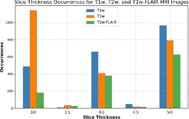
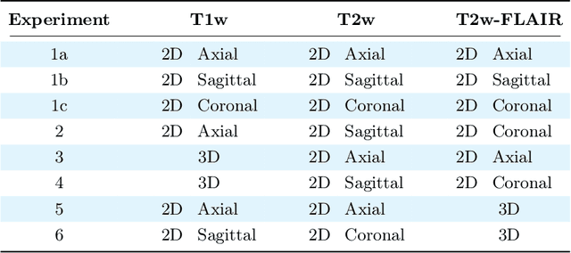
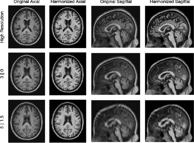

Abstract:Magnetic resonance (MR) imaging is commonly used in the clinical setting to non-invasively monitor the body. There exists a large variability in MR imaging due to differences in scanner hardware, software, and protocol design. Ideally, a processing algorithm should perform robustly to this variability, but that is not always the case in reality. This introduces a need for image harmonization to overcome issues of domain shift when performing downstream analysis such as segmentation. Most image harmonization models focus on acquisition parameters such as inversion time or repetition time, but they ignore an important aspect in MR imaging -- resolution. In this paper, we evaluate the impact of image resolution on harmonization using a pretrained harmonization algorithm. We simulate 2D acquisitions of various slice thicknesses and gaps from 3D acquired, 1mm3 isotropic MR images and demonstrate how the performance of a state-of-the-art image harmonization algorithm varies as resolution changes. We discuss the most ideal scenarios for image resolution including acquisition orientation when 3D imaging is not available, which is common for many clinical scanners. Our results show that harmonization on low-resolution images does not account for acquisition resolution and orientation variations. Super-resolution can be used to alleviate resolution variations but it is not always used. Our methodology can generalize to help evaluate the impact of image acquisition resolution for multiple tasks. Determining the limits of a pretrained algorithm is important when considering preprocessing steps and trust in the results.
HACA3: A Unified Approach for Multi-site MR Image Harmonization
Dec 12, 2022Abstract:The lack of standardization is a prominent issue in magnetic resonance (MR) imaging. This often causes undesired contrast variations due to differences in hardware and acquisition parameters. In recent years, MR harmonization using image synthesis with disentanglement has been proposed to compensate for the undesired contrast variations. Despite the success of existing methods, we argue that three major improvements can be made. First, most existing methods are built upon the assumption that multi-contrast MR images of the same subject share the same anatomy. This assumption is questionable since different MR contrasts are specialized to highlight different anatomical features. Second, these methods often require a fixed set of MR contrasts for training (e.g., both Tw-weighted and T2-weighted images must be available), which limits their applicability. Third, existing methods generally are sensitive to imaging artifacts. In this paper, we present a novel approach, Harmonization with Attention-based Contrast, Anatomy, and Artifact Awareness (HACA3), to address these three issues. We first propose an anatomy fusion module that enables HACA3 to respect the anatomical differences between MR contrasts. HACA3 is also robust to imaging artifacts and can be trained and applied to any set of MR contrasts. Experiments show that HACA3 achieves state-of-the-art performance under multiple image quality metrics. We also demonstrate the applicability of HACA3 on downstream tasks with diverse MR datasets acquired from 21 sites with different field strengths, scanner platforms, and acquisition protocols.
 Add to Chrome
 Add to Firefox
 Add to Edge