Brian Zenger
Interpretable Modeling and Reduction of Unknown Errors in Mechanistic Operators
Nov 02, 2022Abstract:Prior knowledge about the imaging physics provides a mechanistic forward operator that plays an important role in image reconstruction, although myriad sources of possible errors in the operator could negatively impact the reconstruction solutions. In this work, we propose to embed the traditional mechanistic forward operator inside a neural function, and focus on modeling and correcting its unknown errors in an interpretable manner. This is achieved by a conditional generative model that transforms a given mechanistic operator with unknown errors, arising from a latent space of self-organizing clusters of potential sources of error generation. Once learned, the generative model can be used in place of a fixed forward operator in any traditional optimization-based reconstruction process where, together with the inverse solution, the error in prior mechanistic forward operator can be minimized and the potential source of error uncovered. We apply the presented method to the reconstruction of heart electrical potential from body surface potential. In controlled simulation experiments and in-vivo real data experiments, we demonstrate that the presented method allowed reduction of errors in the physics-based forward operator and thereby delivered inverse reconstruction of heart-surface potential with increased accuracy.
* 11 pages, Conference: Medical Image Computing and Computer Assisted Intervention
Few-shot Generation of Personalized Neural Surrogates for Cardiac Simulation via Bayesian Meta-Learning
Oct 06, 2022Abstract:Clinical adoption of personalized virtual heart simulations faces challenges in model personalization and expensive computation. While an ideal solution is an efficient neural surrogate that at the same time is personalized to an individual subject, the state-of-the-art is either concerned with personalizing an expensive simulation model, or learning an efficient yet generic surrogate. This paper presents a completely new concept to achieve personalized neural surrogates in a single coherent framework of meta-learning (metaPNS). Instead of learning a single neural surrogate, we pursue the process of learning a personalized neural surrogate using a small amount of context data from a subject, in a novel formulation of few-shot generative modeling underpinned by: 1) a set-conditioned neural surrogate for cardiac simulation that, conditioned on subject-specific context data, learns to generate query simulations not included in the context set, and 2) a meta-model of amortized variational inference that learns to condition the neural surrogate via simple feed-forward embedding of context data. As test time, metaPNS delivers a personalized neural surrogate by fast feed-forward embedding of a small and flexible number of data available from an individual, achieving -- for the first time -- personalization and surrogate construction for expensive simulations in one end-to-end learning framework. Synthetic and real-data experiments demonstrated that metaPNS was able to improve personalization and predictive accuracy in comparison to conventionally-optimized cardiac simulation models, at a fraction of computation.
Statistical Shape Modeling of Biventricular Anatomy with Shared Boundaries
Sep 12, 2022


Abstract:Statistical shape modeling (SSM) is a valuable and powerful tool to generate a detailed representation of complex anatomy that enables quantitative analysis and the comparison of shapes and their variations. SSM applies mathematics, statistics, and computing to parse the shape into a quantitative representation (such as correspondence points or landmarks) that will help answer various questions about the anatomical variations across the population. Complex anatomical structures have many diverse parts with varying interactions or intricate architecture. For example, the heart is four-chambered anatomy with several shared boundaries between chambers. Coordinated and efficient contraction of the chambers of the heart is necessary to adequately perfuse end organs throughout the body. Subtle shape changes within these shared boundaries of the heart can indicate potential pathological changes that lead to uncoordinated contraction and poor end-organ perfusion. Early detection and robust quantification could provide insight into ideal treatment techniques and intervention timing. However, existing SSM approaches fall short of explicitly modeling the statistics of shared boundaries. This paper presents a general and flexible data-driven approach for building statistical shape models of multi-organ anatomies with shared boundaries that capture morphological and alignment changes of individual anatomies and their shared boundary surfaces throughout the population. We demonstrate the effectiveness of the proposed methods using a biventricular heart dataset by developing shape models that consistently parameterize the cardiac biventricular structure and the interventricular septum (shared boundary surface) across the population data.
Reducing Line-of-block Artifacts in Cardiac Activation Maps Estimated Using ECG Imaging: A Comparison of Source Models and Estimation Methods
Aug 18, 2021


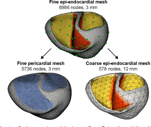
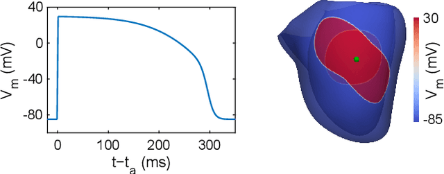
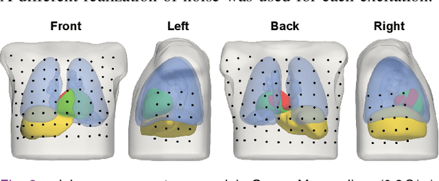
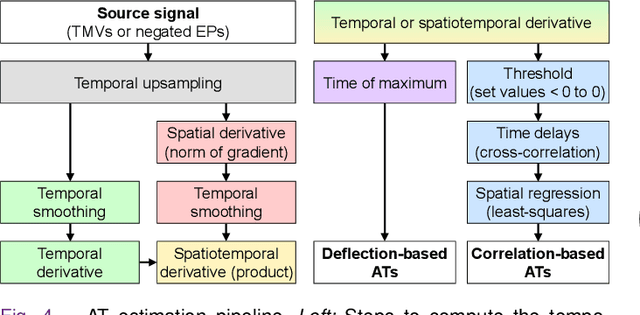
Abstract:Objective: To investigate cardiac activation maps estimated using electrocardiographic imaging and to find methods reducing line-of-block (LoB) artifacts, while preserving real LoBs. Methods: Body surface potentials were computed for 137 simulated ventricular excitations. Subsequently, the inverse problem was solved to obtain extracellular potentials (EP) and transmembrane voltages (TMV). From these, activation times (AT) were estimated using four methods and compared to the ground truth. This process was evaluated with two cardiac mesh resolutions. Factors contributing to LoB artifacts were identified by analyzing the impact of spatial and temporal smoothing on the morphology of source signals. Results: AT estimation using a spatiotemporal derivative performed better than using a temporal derivative. Compared to deflection-based AT estimation, correlation-based methods were less prone to LoB artifacts but performed worse in identifying real LoBs. Temporal smoothing could eliminate artifacts for TMVs but not for EPs, which could be linked to their temporal morphology. TMVs led to more accurate ATs on the septum than EPs. Mesh resolution had a negligible effect on inverse reconstructions, but small distances were important for cross-correlation-based estimation of AT delays. Conclusion: LoB artifacts are mainly caused by the inherent spatial smoothing effect of the inverse reconstruction. Among the configurations evaluated, only deflection-based AT estimation in combination with TMVs and strong temporal smoothing can prevent LoB artifacts, while preserving real LoBs. Significance: Regions of slow conduction are of considerable clinical interest and LoB artifacts observed in non-invasive ATs can lead to misinterpretations. We addressed this problem by identifying factors causing such artifacts and methods to reduce them.
 Add to Chrome
 Add to Firefox
 Add to Edge