Bingjiang Qiu
CKD-TransBTS: Clinical Knowledge-Driven Hybrid Transformer with Modality-Correlated Cross-Attention for Brain Tumor Segmentation
Jul 15, 2022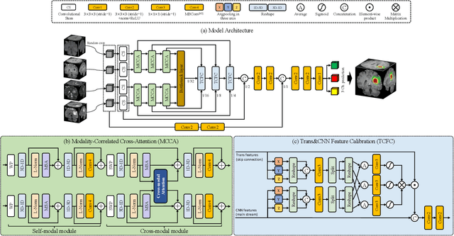
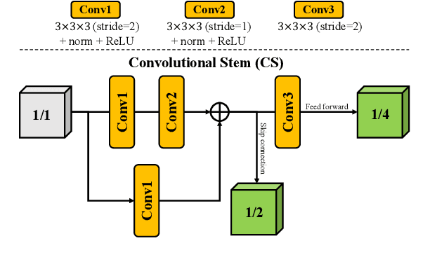
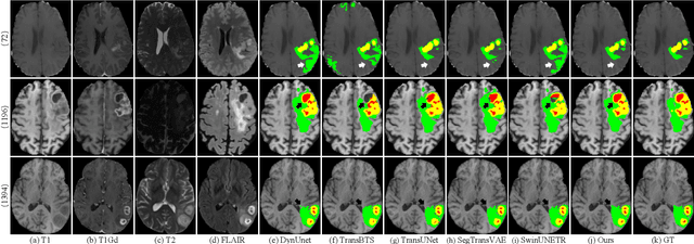
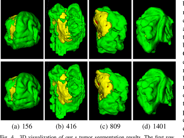
Abstract:Brain tumor segmentation (BTS) in magnetic resonance image (MRI) is crucial for brain tumor diagnosis, cancer management and research purposes. With the great success of the ten-year BraTS challenges as well as the advances of CNN and Transformer algorithms, a lot of outstanding BTS models have been proposed to tackle the difficulties of BTS in different technical aspects. However, existing studies hardly consider how to fuse the multi-modality images in a reasonable manner. In this paper, we leverage the clinical knowledge of how radiologists diagnose brain tumors from multiple MRI modalities and propose a clinical knowledge-driven brain tumor segmentation model, called CKD-TransBTS. Instead of directly concatenating all the modalities, we re-organize the input modalities by separating them into two groups according to the imaging principle of MRI. A dual-branch hybrid encoder with the proposed modality-correlated cross-attention block (MCCA) is designed to extract the multi-modality image features. The proposed model inherits the strengths from both Transformer and CNN with the local feature representation ability for precise lesion boundaries and long-range feature extraction for 3D volumetric images. To bridge the gap between Transformer and CNN features, we propose a Trans&CNN Feature Calibration block (TCFC) in the decoder. We compare the proposed model with five CNN-based models and six transformer-based models on the BraTS 2021 challenge dataset. Extensive experiments demonstrate that the proposed model achieves state-of-the-art brain tumor segmentation performance compared with all the competitors.
HoVer-Trans: Anatomy-aware HoVer-Transformer for ROI-free Breast Cancer Diagnosis in Ultrasound Images
May 17, 2022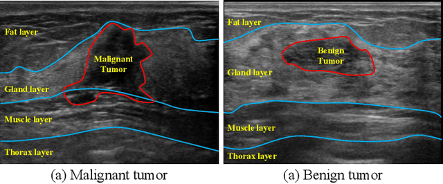
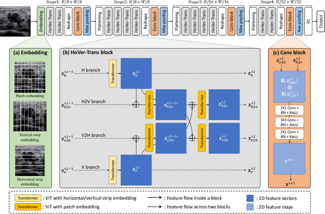
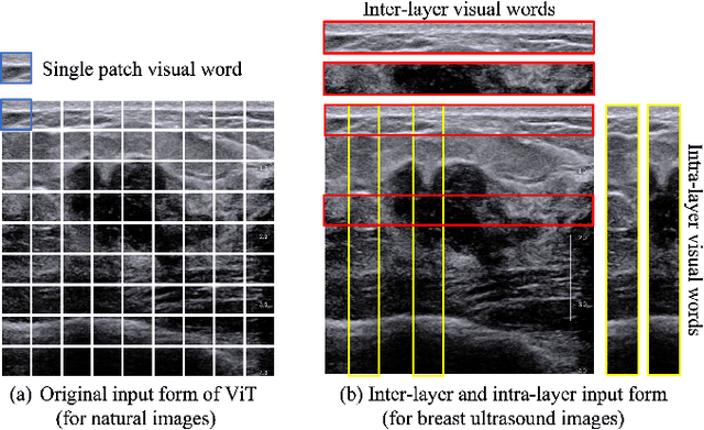
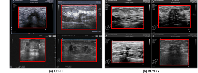
Abstract:Ultrasonography is an important routine examination for breast cancer diagnosis, due to its non-invasive, radiation-free and low-cost properties. However, it is still not the first-line screening test for breast cancer due to its inherent limitations. It would be a tremendous success if we can precisely diagnose breast cancer by breast ultrasound images (BUS). Many learning-based computer-aided diagnostic methods have been proposed to achieve breast cancer diagnosis/lesion classification. However, most of them require a pre-define ROI and then classify the lesion inside the ROI. Conventional classification backbones, such as VGG16 and ResNet50, can achieve promising classification results with no ROI requirement. But these models lack interpretability, thus restricting their use in clinical practice. In this study, we propose a novel ROI-free model for breast cancer diagnosis in ultrasound images with interpretable feature representations. We leverage the anatomical prior knowledge that malignant and benign tumors have different spatial relationships between different tissue layers, and propose a HoVer-Transformer to formulate this prior knowledge. The proposed HoVer-Trans block extracts the inter- and intra-layer spatial information horizontally and vertically. We conduct and release an open dataset GDPH&GYFYY for breast cancer diagnosis in BUS. The proposed model is evaluated in three datasets by comparing with four CNN-based models and two vision transformer models via a five-fold cross validation. It achieves state-of-the-art classification performance with the best model interpretability.
Recurrent convolutional neural networks for mandible segmentation from computed tomography
Mar 13, 2020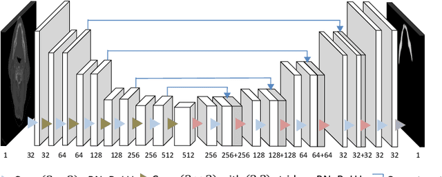
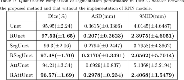
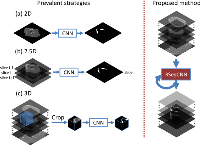

Abstract:Recently, accurate mandible segmentation in CT scans based on deep learning methods has attracted much attention. However, there still exist two major challenges, namely, metal artifacts among mandibles and large variations in shape or size among individuals. To address these two challenges, we propose a recurrent segmentation convolutional neural network (RSegCNN) that embeds segmentation convolutional neural network (SegCNN) into the recurrent neural network (RNN) for robust and accurate segmentation of the mandible. Such a design of the system takes into account the similarity and continuity of the mandible shapes captured in adjacent image slices in CT scans. The RSegCNN infers the mandible information based on the recurrent structure with the embedded encoder-decoder segmentation (SegCNN) components. The recurrent structure guides the system to exploit relevant and important information from adjacent slices, while the SegCNN component focuses on the mandible shapes from a single CT slice. We conducted extensive experiments to evaluate the proposed RSegCNN on two head and neck CT datasets. The experimental results show that the RSegCNN is significantly better than the state-of-the-art models for accurate mandible segmentation.
3D segmentation of mandible from multisectional CT scans by convolutional neural networks
Sep 18, 2018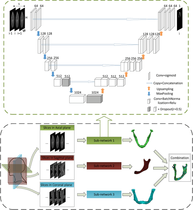
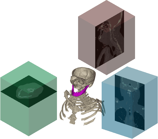


Abstract:Segmentation of mandibles in CT scans during virtual surgical planning is crucial for 3D surgical planning in order to obtain a detailed surface representation of the patients bone. Automatic segmentation of mandibles in CT scans is a challenging task due to large variation in their shape and size between individuals. In order to address this challenge we propose a convolutional neural network approach for mandible segmentation in CT scans by considering the continuum of anatomical structures through different planes. The proposed convolutional neural network adopts the architecture of the U-Net and then combines the resulting 2D segmentations from three different planes into a 3D segmentation. We implement such a segmentation approach on 11 neck CT scans and then evaluate the performance. We achieve an average dice coefficient of $ 0.89 $ on two testing mandible segmentation. Experimental results show that our proposed approach for mandible segmentation in CT scans exhibits high accuracy.
 Add to Chrome
 Add to Firefox
 Add to Edge