Jiapan Guo
Deep learning-based neurodevelopmental assessment in preterm infants
Jan 17, 2026Abstract:Preterm infants (born between 28 and 37 weeks of gestation) face elevated risks of neurodevelopmental delays, making early identification crucial for timely intervention. While deep learning-based volumetric segmentation of brain MRI scans offers a promising avenue for assessing neonatal neurodevelopment, achieving accurate segmentation of white matter (WM) and gray matter (GM) in preterm infants remains challenging due to their comparable signal intensities (isointense appearance) on MRI during early brain development. To address this, we propose a novel segmentation neural network, named Hierarchical Dense Attention Network. Our architecture incorporates a 3D spatial-channel attention mechanism combined with an attention-guided dense upsampling strategy to enhance feature discrimination in low-contrast volumetric data. Quantitative experiments demonstrate that our method achieves superior segmentation performance compared to state-of-the-art baselines, effectively tackling the challenge of isointense tissue differentiation. Furthermore, application of our algorithm confirms that WM and GM volumes in preterm infants are significantly lower than those in term infants, providing additional imaging evidence of the neurodevelopmental delays associated with preterm birth. The code is available at: https://github.com/ICL-SUST/HDAN.
Feature Complementation Architecture for Visual Place Recognition
Jun 14, 2025Abstract:Visual place recognition (VPR) plays a crucial role in robotic localization and navigation. The key challenge lies in constructing feature representations that are robust to environmental changes. Existing methods typically adopt convolutional neural networks (CNNs) or vision Transformers (ViTs) as feature extractors. However, these architectures excel in different aspects -- CNNs are effective at capturing local details. At the same time, ViTs are better suited for modeling global context, making it difficult to leverage the strengths of both. To address this issue, we propose a local-global feature complementation network (LGCN) for VPR which integrates a parallel CNN-ViT hybrid architecture with a dynamic feature fusion module (DFM). The DFM performs dynamic feature fusion through joint modeling of spatial and channel-wise dependencies. Furthermore, to enhance the expressiveness and adaptability of the ViT branch for VPR tasks, we introduce lightweight frequency-to-spatial fusion adapters into the frozen ViT backbone. These adapters enable task-specific adaptation with controlled parameter overhead. Extensive experiments on multiple VPR benchmark datasets demonstrate that the proposed LGCN consistently outperforms existing approaches in terms of localization accuracy and robustness, validating its effectiveness and generalizability.
Second-order Anisotropic Gaussian Directional Derivative Filters for Blob Detection
Apr 30, 2023


Abstract:Interest point detection methods have received increasing attention and are widely used in computer vision tasks such as image retrieval and 3D reconstruction. In this work, second-order anisotropic Gaussian directional derivative filters with multiple scales are used to smooth the input image and a novel blob detection method is proposed. Extensive experiments demonstrate the superiority of our proposed method over state-of-the-art benchmarks in terms of detection performance and robustness to affine transformations.
Recurrent convolutional neural networks for mandible segmentation from computed tomography
Mar 13, 2020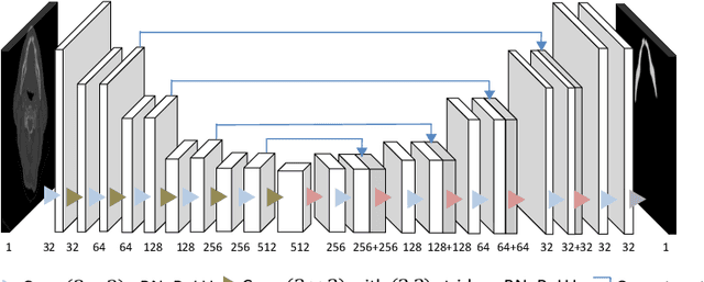
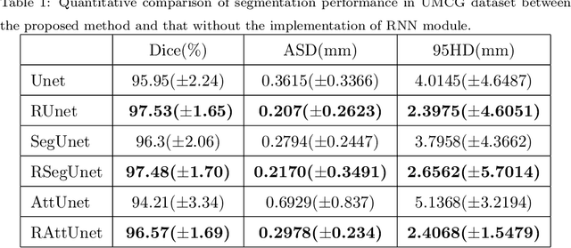
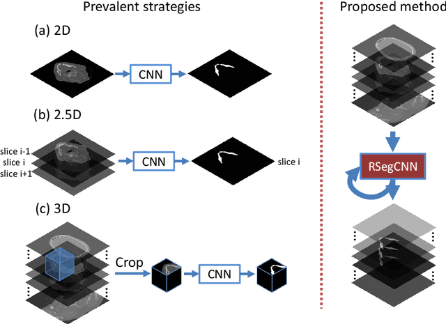

Abstract:Recently, accurate mandible segmentation in CT scans based on deep learning methods has attracted much attention. However, there still exist two major challenges, namely, metal artifacts among mandibles and large variations in shape or size among individuals. To address these two challenges, we propose a recurrent segmentation convolutional neural network (RSegCNN) that embeds segmentation convolutional neural network (SegCNN) into the recurrent neural network (RNN) for robust and accurate segmentation of the mandible. Such a design of the system takes into account the similarity and continuity of the mandible shapes captured in adjacent image slices in CT scans. The RSegCNN infers the mandible information based on the recurrent structure with the embedded encoder-decoder segmentation (SegCNN) components. The recurrent structure guides the system to exploit relevant and important information from adjacent slices, while the SegCNN component focuses on the mandible shapes from a single CT slice. We conducted extensive experiments to evaluate the proposed RSegCNN on two head and neck CT datasets. The experimental results show that the RSegCNN is significantly better than the state-of-the-art models for accurate mandible segmentation.
Automatic Pulmonary Nodule Detection in CT Scans Using Convolutional Neural Networks Based on Maximum Intensity Projection
Apr 11, 2019


Abstract:Accurate pulmonary nodule detection in computed tomography scans is a crucial step in lung cancer screening. Computer-aided detection (CAD) systems are not routinely used by radiologists for pulmonary nodules detection in clinical practice despite their potential benefits. Maximum intensity projection (MIP) images improve the detection of pulmonary nodules in radiological evaluation with computed tomography (CT) scans. In this work, we aim to explore the feasibility of utilizing MIP images to improve the effectiveness of automatic detection of lung nodules by convolutional neural networks (CNNs). We propose a CNN based approach that takes MIP images of different slab thicknesses (5 mm, 10 mm, 15 mm) and 1mm plain multiplanar reconstruction (MPR) images as input. Such an approach augments the 2-D CT slice images with more representative spatial information that helps in the discriminating nodules from vessels through their morphologies. We use the public available LUNA16 set collected from seven academic centers to train and test our approach. Our proposed method achieves a sensitivity of 91.13% with 1 false positive per scan and a sensitivity of 94.13% with 4 false positives per scan for lung nodule detection in this dataset. Using the thick MIP images helps the detection of small pulmonary nodules (3mm-10mm) and acquires fewer false positives. Experimental results show that applying MIP images can increase the sensitivity and lower the number of false positive, which demonstrates the effectiveness and significance of the proposed maximum intensity projection based CNN framework for automatic pulmonary nodule detection in CT scans. Index Terms: Computer-aided detection (CAD), convolutional neural networks (CNNs), computed tomography scans, maximum intensity projection (MIP), pulmonary nodule detection
3D segmentation of mandible from multisectional CT scans by convolutional neural networks
Sep 18, 2018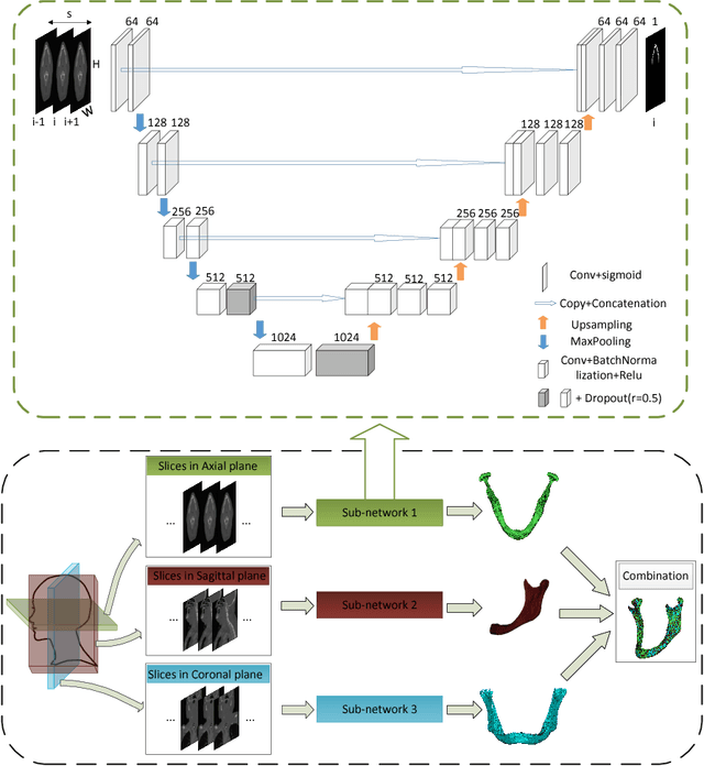
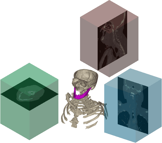


Abstract:Segmentation of mandibles in CT scans during virtual surgical planning is crucial for 3D surgical planning in order to obtain a detailed surface representation of the patients bone. Automatic segmentation of mandibles in CT scans is a challenging task due to large variation in their shape and size between individuals. In order to address this challenge we propose a convolutional neural network approach for mandible segmentation in CT scans by considering the continuum of anatomical structures through different planes. The proposed convolutional neural network adopts the architecture of the U-Net and then combines the resulting 2D segmentations from three different planes into a 3D segmentation. We implement such a segmentation approach on 11 neck CT scans and then evaluate the performance. We achieve an average dice coefficient of $ 0.89 $ on two testing mandible segmentation. Experimental results show that our proposed approach for mandible segmentation in CT scans exhibits high accuracy.
 Add to Chrome
 Add to Firefox
 Add to Edge