Balpreet Singh Ahluwalia
Deep Optical Coding Design in Computational Imaging
Jun 27, 2022


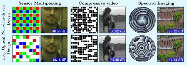
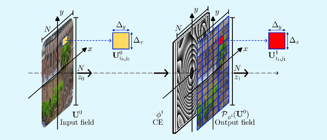
Abstract:Computational optical imaging (COI) systems leverage optical coding elements (CE) in their setups to encode a high-dimensional scene in a single or multiple snapshots and decode it by using computational algorithms. The performance of COI systems highly depends on the design of its main components: the CE pattern and the computational method used to perform a given task. Conventional approaches rely on random patterns or analytical designs to set the distribution of the CE. However, the available data and algorithm capabilities of deep neural networks (DNNs) have opened a new horizon in CE data-driven designs that jointly consider the optical encoder and computational decoder. Specifically, by modeling the COI measurements through a fully differentiable image formation model that considers the physics-based propagation of light and its interaction with the CEs, the parameters that define the CE and the computational decoder can be optimized in an end-to-end (E2E) manner. Moreover, by optimizing just CEs in the same framework, inference tasks can be performed from pure optics. This work surveys the recent advances on CE data-driven design and provides guidelines on how to parametrize different optical elements to include them in the E2E framework. Since the E2E framework can handle different inference applications by changing the loss function and the DNN, we present low-level tasks such as spectral imaging reconstruction or high-level tasks such as pose estimation with privacy preserving enhanced by using optimal task-based optical architectures. Finally, we illustrate classification and 3D object recognition applications performed at the speed of the light using all-optics DNN.
Simulation-supervised deep learning for analysing organelles states and behaviour in living cells
Aug 26, 2020


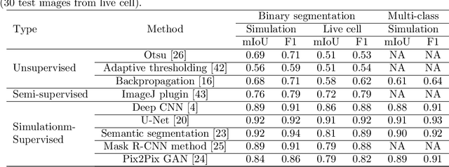
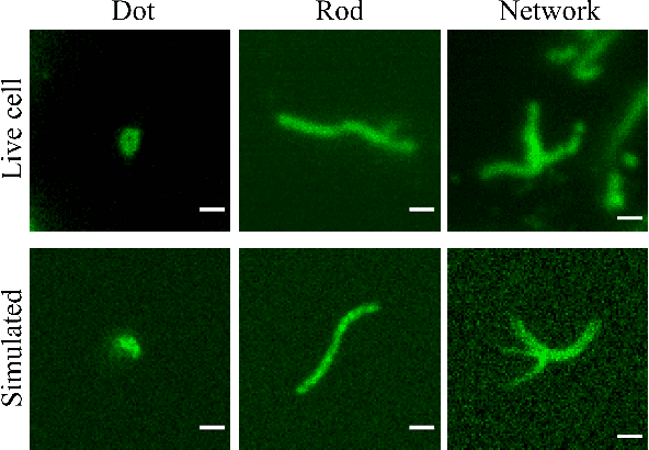
Abstract:In many real-world scientific problems, generating ground truth (GT) for supervised learning is almost impossible. The causes include limitations imposed by scientific instrument, physical phenomenon itself, or the complexity of modeling. Performing artificial intelligence (AI) tasks such as segmentation, tracking, and analytics of small sub-cellular structures such as mitochondria in microscopy videos of living cells is a prime example. The 3D blurring function of microscope, digital resolution from pixel size, optical resolution due to the character of light, noise characteristics, and complex 3D deformable shapes of mitochondria, all contribute to making this problem GT hard. Manual segmentation of 100s of mitochondria across 1000s of frames and then across many such videos is not only herculean but also physically inaccurate because of the instrument and phenomena imposed limitations. Unsupervised learning produces less than optimal results and accuracy is important if inferences relevant to therapy are to be derived. In order to solve this unsurmountable problem, we bring modeling and deep learning to a nexus. We show that accurate physics based modeling of microscopy data including all its limitations can be the solution for generating simulated training datasets for supervised learning. We show here that our simulation-supervised segmentation approach is a great enabler for studying mitochondrial states and behaviour in heart muscle cells, where mitochondria have a significant role to play in the health of the cells. We report unprecedented mean IoU score of 91% for binary segmentation (19% better than the best performing unsupervised approach) of mitochondria in actual microscopy videos of living cells. We further demonstrate the possibility of performing multi-class classification, tracking, and morphology associated analytics at the scale of individual mitochondrion.
 Add to Chrome
 Add to Firefox
 Add to Edge