Weihua Liu
Learning High-Quality Initial Noise for Single-View Synthesis with Diffusion Models
Dec 18, 2025Abstract:Single-view novel view synthesis (NVS) models based on diffusion models have recently attracted increasing attention, as they can generate a series of novel view images from a single image prompt and camera pose information as conditions. It has been observed that in diffusion models, certain high-quality initial noise patterns lead to better generation results than others. However, there remains a lack of dedicated learning frameworks that enable NVS models to learn such high-quality noise. To obtain high-quality initial noise from random Gaussian noise, we make the following contributions. First, we design a discretized Euler inversion method to inject image semantic information into random noise, thereby constructing paired datasets of random and high-quality noise. Second, we propose a learning framework based on an encoder-decoder network (EDN) that directly transforms random noise into high-quality noise. Experiments demonstrate that the proposed EDN can be seamlessly plugged into various NVS models, such as SV3D and MV-Adapter, achieving significant performance improvements across multiple datasets. Code is available at: https://github.com/zhihao0512/EDN.
BENet: A Cross-domain Robust Network for Detecting Face Forgeries via Bias Expansion and Latent-space Attention
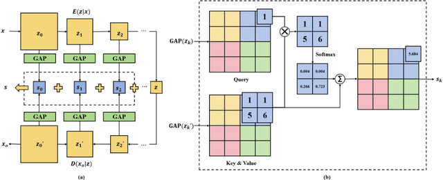
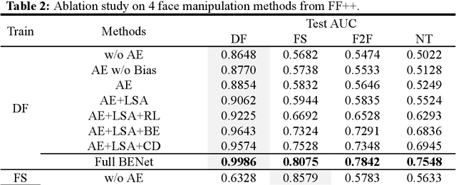
Dec 10, 2024Abstract:In response to the growing threat of deepfake technology, we introduce BENet, a Cross-Domain Robust Bias Expansion Network. BENet enhances the detection of fake faces by addressing limitations in current detectors related to variations across different types of fake face generation techniques, where ``cross-domain" refers to the diverse range of these deepfakes, each considered a separate domain. BENet's core feature is a bias expansion module based on autoencoders. This module maintains genuine facial features while enhancing differences in fake reconstructions, creating a reliable bias for detecting fake faces across various deepfake domains. We also introduce a Latent-Space Attention (LSA) module to capture inconsistencies related to fake faces at different scales, ensuring robust defense against advanced deepfake techniques. The enriched LSA feature maps are multiplied with the expanded bias to create a versatile feature space optimized for subtle forgeries detection. To improve its ability to detect fake faces from unknown sources, BENet integrates a cross-domain detector module that enhances recognition accuracy by verifying the facial domain during inference. We train our network end-to-end with a novel bias expansion loss, adopted for the first time, in face forgery detection. Extensive experiments covering both intra and cross-dataset demonstrate BENet's superiority over current state-of-the-art solutions.
Reconstructing Deep Neural Networks: Unleashing the Optimization Potential of Natural Gradient Descent
Dec 10, 2024


Abstract:Natural gradient descent (NGD) is a powerful optimization technique for machine learning, but the computational complexity of the inverse Fisher information matrix limits its application in training deep neural networks. To overcome this challenge, we propose a novel optimization method for training deep neural networks called structured natural gradient descent (SNGD). Theoretically, we demonstrate that optimizing the original network using NGD is equivalent to using fast gradient descent (GD) to optimize the reconstructed network with a structural transformation of the parameter matrix. Thereby, we decompose the calculation of the global Fisher information matrix into the efficient computation of local Fisher matrices via constructing local Fisher layers in the reconstructed network to speed up the training. Experimental results on various deep networks and datasets demonstrate that SNGD achieves faster convergence speed than NGD while retaining comparable solutions. Furthermore, our method outperforms traditional GDs in terms of efficiency and effectiveness. Thus, our proposed method has the potential to significantly improve the scalability and efficiency of NGD in deep learning applications. Our source code is available at https://github.com/Chaochao-Lin/SNGD.
Cross-domain Robust Deepfake Bias Expansion Network for Face Forgery Detection
Oct 08, 2023


Abstract:The rapid advancement of deepfake technologies raises significant concerns about the security of face recognition systems. While existing methods leverage the clues left by deepfake techniques for face forgery detection, malicious users may intentionally manipulate forged faces to obscure the traces of deepfake clues and thereby deceive detection tools. Meanwhile, attaining cross-domain robustness for data-based methods poses a challenge due to potential gaps in the training data, which may not encompass samples from all relevant domains. Therefore, in this paper, we introduce a solution - a Cross-Domain Robust Bias Expansion Network (BENet) - designed to enhance face forgery detection. BENet employs an auto-encoder to reconstruct input faces, maintaining the invariance of real faces while selectively enhancing the difference between reconstructed fake faces and their original counterparts. This enhanced bias forms a robust foundation upon which dependable forgery detection can be built. To optimize the reconstruction results in BENet, we employ a bias expansion loss infused with contrastive concepts to attain the aforementioned objective. In addition, to further heighten the amplification of forged clues, BENet incorporates a Latent-Space Attention (LSA) module. This LSA module effectively captures variances in latent features between the auto-encoder's encoder and decoder, placing emphasis on inconsistent forgery-related information. Furthermore, BENet incorporates a cross-domain detector with a threshold to determine whether the sample belongs to a known distribution. The correction of classification results through the cross-domain detector enables BENet to defend against unknown deepfake attacks from cross-domain. Extensive experiments demonstrate the superiority of BENet compared with state-of-the-art methods in intra-database and cross-database evaluations.
Dynamic Multi-Domain Knowledge Networks for Chest X-ray Report Generation
Oct 08, 2023Abstract:The automated generation of radiology diagnostic reports helps radiologists make timely and accurate diagnostic decisions while also enhancing clinical diagnostic efficiency. However, the significant imbalance in the distribution of data between normal and abnormal samples (including visual and textual biases) poses significant challenges for a data-driven task like automatically generating diagnostic radiology reports. Therefore, we propose a Dynamic Multi-Domain Knowledge(DMDK) network for radiology diagnostic report generation. The DMDK network consists of four modules: Chest Feature Extractor(CFE), Dynamic Knowledge Extractor(DKE), Specific Knowledge Extractor(SKE), and Multi-knowledge Integrator(MKI) module. Specifically, the CFE module is primarily responsible for extracting the unprocessed visual medical features of the images. The DKE module is responsible for extracting dynamic disease topic labels from the retrieved radiology diagnostic reports. We then fuse the dynamic disease topic labels with the original visual features of the images to highlight the abnormal regions in the original visual features to alleviate the visual data bias problem. The SKE module expands upon the conventional static knowledge graph to mitigate textual data biases and amplify the interpretability capabilities of the model via domain-specific dynamic knowledge graphs. The MKI distills all the knowledge and generates the final diagnostic radiology report. We performed extensive experiments on two widely used datasets, IU X-Ray and MIMIC-CXR. The experimental results demonstrate the effectiveness of our method, with all evaluation metrics outperforming previous state-of-the-art models.
Identifying Distribution Network Faults Using Adaptive Transition Probability
Sep 30, 2023Abstract:A novel approach is suggested for improving the accuracy of fault detection in distribution networks. This technique combines adaptive probability learning and waveform decomposition to optimize the similarity of features. Its objective is to discover the most appropriate linear mapping between simulated and real data to minimize distribution differences. By aligning the data in the same feature space, the proposed method effectively overcomes the challenge posed by limited sample size when identifying faults and classifying real data in distribution networks. Experimental results utilizing simulated system data and real field data demonstrate that this approach outperforms commonly used classification models such as convolutional neural networks, support vector machines, and k-nearest neighbors, especially under adaptive learning conditions. Consequently, this research provides a fresh perspective on fault detection in distribution networks, particularly when adaptive learning conditions are employed.
Shape-Margin Knowledge Augmented Network for Thyroid Nodule Segmentation and Diagnosis
Aug 29, 2023


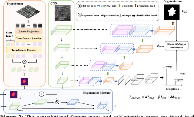
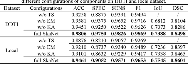
Abstract:Thyroid nodule segmentation is a crucial step in the diagnostic procedure of physicians and computer-aided diagnosis systems. Mostly, current studies treat segmentation and diagnosis as independent tasks without considering the correlation between these tasks. The sequence steps of these independent tasks in computer-aided diagnosis systems may lead to the accumulation of errors. Therefore, it is worth combining them as a whole through exploring the relationship between thyroid nodule segmentation and diagnosis. According to the thyroid imaging reporting and data system (TI-RADS), the assessment of shape and margin characteristics is the prerequisite for the discrimination of benign and malignant thyroid nodules. These characteristics can be observed in the thyroid nodule segmentation masks. Inspired by the diagnostic procedure of TI-RADS, this paper proposes a shape-margin knowledge augmented network (SkaNet) for simultaneously thyroid nodule segmentation and diagnosis. Due to the similarity in visual features between segmentation and diagnosis, SkaNet shares visual features in the feature extraction stage and then utilizes a dual-branch architecture to perform thyroid nodule segmentation and diagnosis tasks simultaneously. To enhance effective discriminative features, an exponential mixture module is devised, which incorporates convolutional feature maps and self-attention maps by exponential weighting. Then, SkaNet is jointly optimized by a knowledge augmented multi-task loss function with a constraint penalty term. It embeds shape and margin characteristics through numerical computation and models the relationship between the thyroid nodule diagnosis results and segmentation masks.
Occlusion-Aware Deep Convolutional Neural Network via Homogeneous Tanh-transforms for Face Parsing
Aug 29, 2023Abstract:Face parsing infers a pixel-wise label map for each semantic facial component. Previous methods generally work well for uncovered faces, however overlook the facial occlusion and ignore some contextual area outside a single face, especially when facial occlusion has become a common situation during the COVID-19 epidemic. Inspired by the illumination theory of image, we propose a novel homogeneous tanh-transforms for image preprocessing, which made up of four tanh-transforms, that fuse the central vision and the peripheral vision together. Our proposed method addresses the dilemma of face parsing under occlusion and compresses more information of surrounding context. Based on homogeneous tanh-transforms, we propose an occlusion-aware convolutional neural network for occluded face parsing. It combines the information both in Tanh-polar space and Tanh-Cartesian space, capable of enhancing receptive fields. Furthermore, we introduce an occlusion-aware loss to focus on the boundaries of occluded regions. The network is simple and flexible, and can be trained end-to-end. To facilitate future research of occluded face parsing, we also contribute a new cleaned face parsing dataset, which is manually purified from several academic or industrial datasets, including CelebAMask-HQ, Short-video Face Parsing as well as Helen dataset and will make it public. Experiments demonstrate that our method surpasses state-of-art methods of face parsing under occlusion.
Enhancing Mobile Face Anti-Spoofing: A Robust Framework for Diverse Attack Types under Screen Flash
Aug 29, 2023Abstract:Face anti-spoofing (FAS) is crucial for securing face recognition systems. However, existing FAS methods with handcrafted binary or pixel-wise labels have limitations due to diverse presentation attacks (PAs). In this paper, we propose an attack type robust face anti-spoofing framework under light flash, called ATR-FAS. Due to imaging differences caused by various attack types, traditional FAS methods based on single binary classification network may result in excessive intra-class distance of spoof faces, leading to a challenge of decision boundary learning. Therefore, we employed multiple networks to reconstruct multi-frame depth maps as auxiliary supervision, and each network experts in one type of attack. A dual gate module (DGM) consisting of a type gate and a frame-attention gate is introduced, which perform attack type recognition and multi-frame attention generation, respectively. The outputs of DGM are utilized as weight to mix the result of multiple expert networks. The multi-experts mixture enables ATR-FAS to generate spoof-differentiated depth maps, and stably detects spoof faces without being affected by different types of PAs. Moreover, we design a differential normalization procedure to convert original flash frames into differential frames. This simple but effective processing enhances the details in flash frames, aiding in the generation of depth maps. To verify the effectiveness of our framework, we collected a large-scale dataset containing 12,660 live and spoof videos with diverse PAs under dynamic flash from the smartphone screen. Extensive experiments illustrate that the proposed ATR-FAS significantly outperforms existing state-of-the-art methods. The code and dataset will be available at https://github.com/Chaochao-Lin/ATR-FAS.
Stone Needle: A General Multimodal Large-scale Model Framework towards Healthcare
Jun 28, 2023Abstract:In healthcare, multimodal data is prevalent and requires to be comprehensively analyzed before diagnostic decisions, including medical images, clinical reports, etc. However, current large-scale artificial intelligence models predominantly focus on single-modal cognitive abilities and neglect the integration of multiple modalities. Therefore, we propose Stone Needle, a general multimodal large-scale model framework tailored explicitly for healthcare applications. Stone Needle serves as a comprehensive medical multimodal model foundation, integrating various modalities such as text, images, videos, and audio to surpass the limitations of single-modal systems. Through the framework components of intent analysis, medical foundation models, prompt manager, and medical language module, our architecture can perform multi-modal interaction in multiple rounds of dialogue. Our method is a general multimodal large-scale model framework, integrating diverse modalities and allowing us to tailor for specific tasks. The experimental results demonstrate the superior performance of our method compared to single-modal systems. The fusion of different modalities and the ability to process complex medical information in Stone Needle benefits accurate diagnosis, treatment recommendations, and patient care.
 Add to Chrome
 Add to Firefox
 Add to Edge