Vincent Estrade
Deep morphological recognition of kidney stones using intra-operative endoscopic digital videos
May 12, 2022

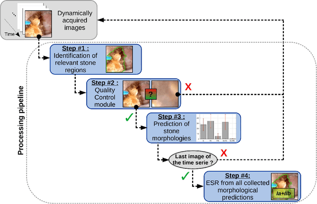
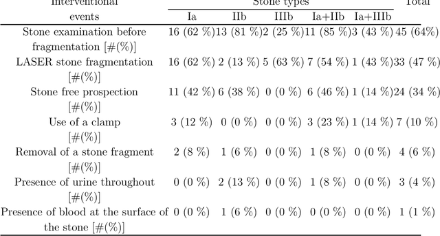
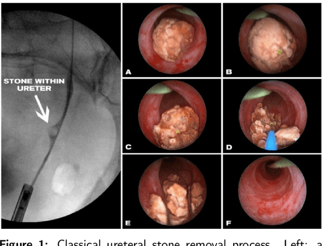
Abstract:The collection and the analysis of kidney stone morphological criteria are essential for an aetiological diagnosis of stone disease. However, in-situ LASER-based fragmentation of urinary stones, which is now the most established chirurgical intervention, may destroy the morphology of the targeted stone. In the current study, we assess the performance and added value of processing complete digital endoscopic video sequences for the automatic recognition of stone morphological features during a standard-of-care intra-operative session. To this end, a computer-aided video classifier was developed to predict in-situ the morphology of stone using an intra-operative digital endoscopic video acquired in a clinical setting. The proposed technique was evaluated on pure (i.e. include one morphology) and mixed (i.e. include at least two morphologies) stones involving "Ia/Calcium Oxalate Monohydrate (COM)", "IIb/ Calcium Oxalate Dihydrate (COD)" and "IIIb/Uric Acid (UA)" morphologies. 71 digital endoscopic videos (50 exhibited only one morphological type and 21 displayed two) were analyzed using the proposed video classifier (56840 frames processed in total). Using the proposed approach, diagnostic performances (averaged over both pure and mixed stone types) were as follows: balanced accuracy=88%, sensitivity=80%, specificity=95%, precision=78% and F1-score=78%. The obtained results demonstrate that AI applied on digital endoscopic video sequences is a promising tool for collecting morphological information during the time-course of the stone fragmentation process without resorting to any human intervention for stone delineation or selection of good quality steady frames. To this end, irrelevant image information must be removed from the prediction process at both frame and pixel levels, which is now feasible thanks to the use of AI-dedicated networks.
On the generalization capabilities of FSL methods through domain adaptation: a case study in endoscopic kidney stone image classification
May 02, 2022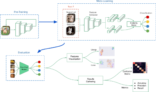


Abstract:Deep learning has shown great promise in diverse areas of computer vision, such as image classification, object detection and semantic segmentation, among many others. However, as it has been repeatedly demonstrated, deep learning methods trained on a dataset do not generalize well to datasets from other domains or even to similar datasets, due to data distribution shifts. In this work, we propose the use of a meta-learning based few-shot learning approach to alleviate these problems. In order to demonstrate its efficacy, we use two datasets of kidney stones samples acquired with different endoscopes and different acquisition conditions. The results show how such methods are indeed capable of handling domain-shifts by attaining an accuracy of 74.38% and 88.52% in the 5-way 5-shot and 5-way 20-shot settings respectively. Instead, in the same dataset, traditional Deep Learning (DL) methods attain only an accuracy of 45%.
On the in vivo recognition of kidney stones using machine learning
Jan 21, 2022

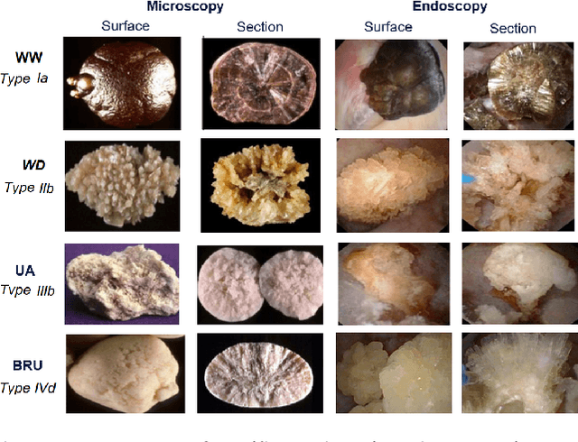
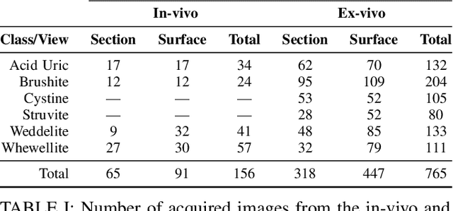
Abstract:Determining the type of kidney stones allows urologists to prescribe a treatment to avoid recurrence of renal lithiasis. An automated in-vivo image-based classification method would be an important step towards an immediate identification of the kidney stone type required as a first phase of the diagnosis. In the literature it was shown on ex-vivo data (i.e., in very controlled scene and image acquisition conditions) that an automated kidney stone classification is indeed feasible. This pilot study compares the kidney stone recognition performances of six shallow machine learning methods and three deep-learning architectures which were tested with in-vivo images of the four most frequent urinary calculi types acquired with an endoscope during standard ureteroscopies. This contribution details the database construction and the design of the tested kidney stones classifiers. Even if the best results were obtained by the Inception v3 architecture (weighted precision, recall and F1-score of 0.97, 0.98 and 0.97, respectively), it is also shown that choosing an appropriate colour space and texture features allows a shallow machine learning method to approach closely the performances of the most promising deep-learning methods (the XGBoost classifier led to weighted precision, recall and F1-score values of 0.96). This paper is the first one that explores the most discriminant features to be extracted from images acquired during ureteroscopies.
Towards Automatic Recognition of Pure & Mixed Stones using Intraoperative Endoscopic Digital Images
May 22, 2021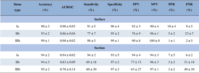
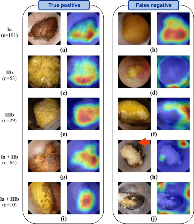
Abstract:Objective: To assess automatic computer-aided in-situ recognition of morphological features of pure and mixed urinary stones using intraoperative digital endoscopic images acquired in a clinical setting. Materials and methods: In this single-centre study, an experienced urologist intraoperatively and prospectively examined the surface and section of all kidney stones encountered. Calcium oxalate monohydrate (COM/Ia), dihydrate (COD/IIb) and uric acid (UA/IIIb) morphological criteria were collected and classified to generate annotated datasets. A deep convolutional neural network (CNN) was trained to predict the composition of both pure and mixed stones. To explain the predictions of the deep neural network model, coarse localisation heat-maps were plotted to pinpoint key areas identified by the network. Results: This study included 347 and 236 observations of stone surface and stone section, respectively. A highest sensitivity of 98 % was obtained for the type "pure IIIb/UA" using surface images. The most frequently encountered morphology was that of the type "pure Ia/COM"; it was correctly predicted in 91 % and 94 % of cases using surface and section images, respectively. Of the mixed type "Ia/COM+IIb/COD", Ia/COM was predicted in 84 % of cases using surface images, IIb/COD in 70 % of cases, and both in 65 % of cases. Concerning mixed Ia/COM+IIIb/UA stones, Ia/COM was predicted in 91 % of cases using section images, IIIb/UA in 69 % of cases, and both in 74 % of cases. Conclusions: This preliminary study demonstrates that deep convolutional neural networks are promising to identify kidney stone composition from endoscopic images acquired intraoperatively. Both pure and mixed stone composition could be discriminated. Collected in a clinical setting, surface and section images analysed by deep CNN provide valuable information about stone morphology for computer-aided diagnosis.
Assessing deep learning methods for the identification of kidney stones in endoscopic images
Mar 01, 2021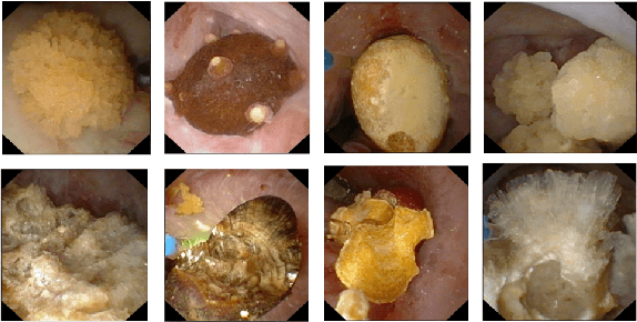
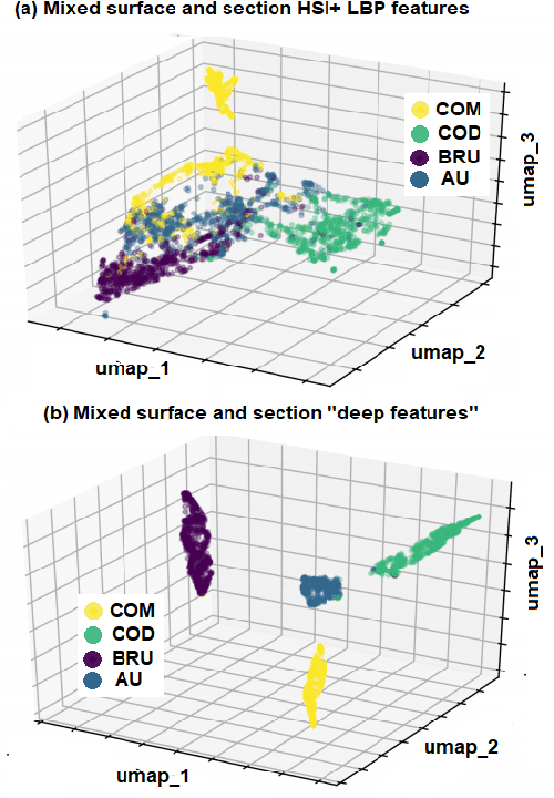

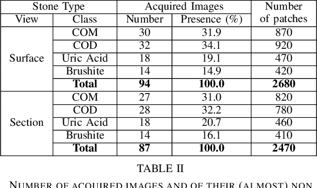
Abstract:Knowing the type (i.e., the biochemical composition) of kidney stones is crucial to prevent relapses with an appropriate treatment. During ureteroscopies, kidney stones are fragmented, extracted from the urinary tract, and their composition is determined using a morpho-constitutional analysis. This procedure is time consuming (the morpho-constitutional analysis results are only available after some days) and tedious (the fragment extraction lasts up to an hour). Identifying the kidney stone type only with the in-vivo endoscopic images would allow for the dusting of the fragments, while the morpho-constitutional analysis could be avoided. Only few contributions dealing with the in vivo identification of kidney stones were published. This paper discusses and compares five classification methods including deep convolutional neural networks (DCNN)-based approaches and traditional (non DCNN-based) ones. Even if the best method is a DCCN approach with a precision and recall of 98% and 97% over four classes, this contribution shows that a XGBoost classifier exploiting well-chosen feature vectors can closely approach the performances of DCNN classifiers for a medical application with a limited number of annotated data.
 Add to Chrome
 Add to Firefox
 Add to Edge