Thanh D. Nguyen
Video-rate gigapixel ptychography via space-time neural field representations
Nov 08, 2025Abstract:Achieving gigapixel space-bandwidth products (SBP) at video rates represents a fundamental challenge in imaging science. Here we demonstrate video-rate ptychography that overcomes this barrier by exploiting spatiotemporal correlations through neural field representations. Our approach factorizes the space-time volume into low-rank spatial and temporal features, transforming SBP scaling from sequential measurements to efficient correlation extraction. The architecture employs dual networks for decoding real and imaginary field components, avoiding phase-wrapping discontinuities plagued in amplitude-phase representations. A gradient-domain loss on spatial derivatives ensures robust convergence. We demonstrate video-rate gigapixel imaging with centimeter-scale coverage while resolving 308-nm linewidths. Validations span from monitoring sample dynamics of crystals, bacteria, stem cells, microneedle to characterizing time-varying probes in extreme ultraviolet experiments, demonstrating versatility across wavelengths. By transforming temporal variations from a constraint into exploitable correlations, we establish that gigapixel video is tractable with single-sensor measurements, making ptychography a high-throughput sensing tool for monitoring mesoscale dynamics without lenses.
Deep-ultraviolet ptychographic pocket-scope (DART): mesoscale lensless molecular imaging with label-free spectroscopic contrast
Nov 08, 2025Abstract:The mesoscale characterization of biological specimens has traditionally required compromises between resolution, field-of-view, depth-of-field, and molecular specificity, with most approaches relying on external labels. Here we present the Deep-ultrAviolet ptychogRaphic pockeT-scope (DART), a handheld platform that transforms label-free molecular imaging through intrinsic deep-ultraviolet spectroscopic contrast. By leveraging biomolecules' natural absorption fingerprints and combining them with lensless ptychographic microscopy, DART resolves down to 308-nm linewidths across centimeter-scale areas while maintaining millimeter-scale depth-of-field. The system's virtual error-bin methodology effectively eliminates artifacts from limited temporal coherence and other optical imperfections, enabling high-fidelity molecular imaging without lenses. Through differential spectroscopic imaging at deep-ultraviolet wavelengths, DART quantitatively maps nucleic acid and protein distributions with femtogram sensitivity, providing an intrinsic basis for explainable virtual staining. We demonstrate DART's capabilities through molecular imaging of tissue sections, cytopathology specimens, blood cells, and neural populations, revealing detailed molecular contrast without external labels. The combination of high-resolution molecular mapping and broad mesoscale imaging in a portable platform opens new possibilities from rapid clinical diagnostics, tissue analysis, to biological characterization in space exploration.
Synthetic Generation and Latent Projection Denoising of Rim Lesions in Multiple Sclerosis
May 29, 2025Abstract:Quantitative susceptibility maps from magnetic resonance images can provide both prognostic and diagnostic information in multiple sclerosis, a neurodegenerative disease characterized by the formation of lesions in white matter brain tissue. In particular, susceptibility maps provide adequate contrast to distinguish between "rim" lesions, surrounded by deposited paramagnetic iron, and "non-rim" lesion types. These paramagnetic rim lesions (PRLs) are an emerging biomarker in multiple sclerosis. Much effort has been devoted to both detection and segmentation of such lesions to monitor longitudinal change. As paramagnetic rim lesions are rare, addressing this problem requires confronting the class imbalance between rim and non-rim lesions. We produce synthetic quantitative susceptibility maps of paramagnetic rim lesions and show that inclusion of such synthetic data improves classifier performance and provide a multi-channel extension to generate accompanying contrasts and probabilistic segmentation maps. We exploit the projection capability of our trained generative network to demonstrate a novel denoising approach that allows us to train on ambiguous rim cases and substantially increase the minority class. We show that both synthetic lesion synthesis and our proposed rim lesion label denoising method best approximate the unseen rim lesion distribution and improve detection in a clinically interpretable manner. We release our code and generated data at https://github.com/agr78/PRLx-GAN upon publication.
Implicit neural representation for free-breathing MR fingerprinting (INR-MRF): co-registered 3D whole-liver water T1, water T2, proton density fat fraction, and R2* mapping
Oct 19, 2024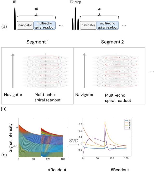

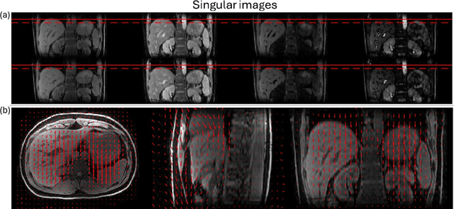
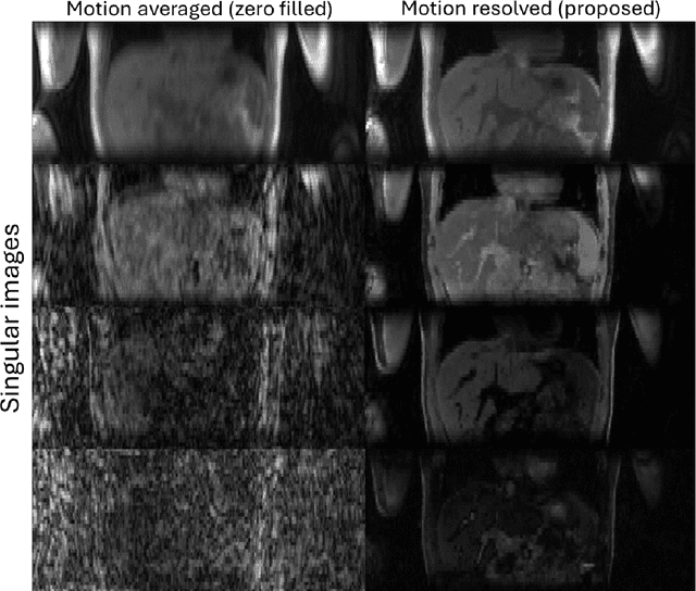
Abstract:Purpose: To develop an MRI technique for free-breathing 3D whole-liver quantification of water T1, water T2, proton density fat fraction (PDFF), R2*. Methods: An Eight-echo spoiled gradient echo pulse sequence with spiral readout was developed by interleaving inversion recovery and T2 magnetization preparation. We propose a neural network based on a 4D and a 3D implicit neural representation (INR) which simultaneously learns the motion deformation fields and the static reference frame MRI subspace images respectively. Water and fat singular images were separated during network training, with no need of performing retrospective water-fat separation. T1, T2, R2* and proton density fat fraction (PDFF) produced by the proposed method were validated in vivo on 10 healthy subjects, using quantitative maps generated from conventional scans as reference. Results: Our results showed minimal bias and narrow 95% limits of agreement on T1, T2, R2* and PDFF values in the liver compared to conventional breath-holding scans. Conclusions: INR-MRF enabled co-registered 3D whole liver T1, T2, R2* and PDFF mapping in a single free-breathing scan.
MRI quantification of liver fibrosis using diamagnetic susceptibility: An ex-vivo feasibility study
Oct 04, 2024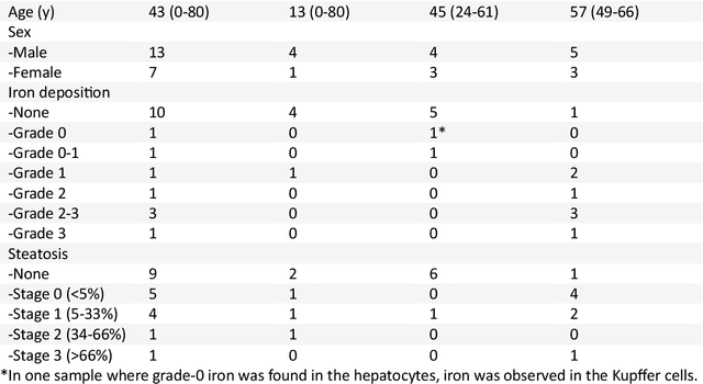
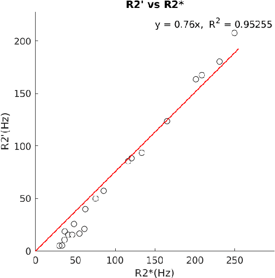
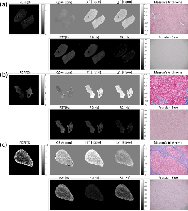

Abstract:In chronic liver disease, liver fibrosis develops as excessive deposition of extracellular matrix macromolecules, predominantly collagens, progressively form fibrous scars that disrupt the hepatic architecture, and fibrosis, iron, and fat are interrelated. Fibrosis is the best predictor of morbidity and mortality in chronic liver disease but liver biopsy, the reference method for diagnosis and staging, is invasive and limited by sampling and interobserver variability and risks of complications. The overall objective of this study was to develop a new non-invasive method to quantify fibrosis using diamagnetic susceptibility sources with histology validation in ex vivo liver explants.
Deep learning improved autofocus for motion artifact reduction and its application in quantitative susceptibility mapping
May 26, 2024Abstract:Purpose: To develop a pipeline for motion artifact correction in mGRE and quantitative susceptibility mapping (QSM). Methods: Deep learning is integrated with autofocus to improve motion artifact suppression, which is applied QSM of patients with Parkinson's disease (PD). The estimation of affine motion parameters in the autofocus method depends on signal-to-noise ratio and lacks accuracy when data sampling occurs outside the k-space center. A deep learning strategy is employed to remove the residual motion artifacts in autofocus. Results: Results obtained in simulated brain data (n =15) with reference truth show that the proposed autofocus deep learning method significantly improves the image quality of mGRE and QSM (p = 0.001 for SSIM, p < 0.0001 for PSNR and RMSE). Results from 10 PD patients with real motion artifacts in QSM have also been corrected using the proposed method and sent to an experienced radiologist for image quality evaluation, and the average image quality score has increased (p=0.0039). Conclusions: The proposed method enables substantial suppression of motion artifacts in mGRE and QSM.
RimSet: Quantitatively Identifying and Characterizing Chronic Active Multiple Sclerosis Lesion on Quantitative Susceptibility Maps
Dec 28, 2023Abstract:Background: Rim+ lesions in multiple sclerosis (MS), detectable via Quantitative Susceptibility Mapping (QSM), correlate with increased disability. Existing literature lacks quantitative analysis of these lesions. We introduce RimSet for quantitative identification and characterization of rim+ lesions on QSM. Methods: RimSet combines RimSeg, an unsupervised segmentation method using level-set methodology, and radiomic measurements with Local Binary Pattern texture descriptors. We validated RimSet using simulated QSM images and an in vivo dataset of 172 MS subjects with 177 rim+ and 3986 rim-lesions. Results: RimSeg achieved a 78.7% Dice score against the ground truth, with challenges in partial rim lesions. RimSet detected rim+ lesions with a partial ROC AUC of 0.808 and PR AUC of 0.737, surpassing existing methods. QSMRim-Net showed the lowest mean square error (0.85) and high correlation (0.91; 95% CI: 0.88, 0.93) with expert annotations at the subject level.
Multi-delay arterial spin-labeled perfusion estimation with biophysics simulation and deep learning
Nov 17, 2023


Abstract:Purpose: To develop biophysics-based method for estimating perfusion Q from arterial spin labeling (ASL) images using deep learning. Methods: A 3D U-Net (QTMnet) was trained to estimate perfusion from 4D tracer propagation images. The network was trained and tested on simulated 4D tracer concentration data based on artificial vasculature structure generated by constrained constructive optimization (CCO) method. The trained network was further tested in a synthetic brain ASL image based on vasculature network extracted from magnetic resonance (MR) angiography. The estimations from both trained network and a conventional kinetic model were compared in ASL images acquired from eight healthy volunteers. Results: QTMnet accurately reconstructed perfusion Q from concentration data. Relative error of the synthetic brain ASL image was 7.04% for perfusion Q, lower than the error using single-delay ASL model: 25.15% for Q, and multi-delay ASL model: 12.62% for perfusion Q. Conclusion: QTMnet provides accurate estimation on perfusion parameters and is a promising approach as a clinical ASL MRI image processing pipeline.
Physics-based network fine-tuning for robust quantitative susceptibility mapping from high-pass filtered phase
May 05, 2023


Abstract:Purpose: To improve the generalization ability of convolutional neural network (CNN) based prediction of quantitative susceptibility mapping (QSM) from high-pass filtered phase (HPFP) image. Methods: The proposed network addresses two common generalization issues that arise when using a pre-trained network to predict QSM from HPFP: a) data with unseen voxel sizes, and b) data with unknown high-pass filter parameters. A network fine-tuning step based on a high-pass filtering dipole convolution forward model is proposed to reduce the generalization error of the pre-trained network. A progressive Unet architecture is proposed to improve prediction accuracy without increasing fine-tuning computational cost. Results: In retrospective studies using RMSE, PSNR, SSIM and HFEN as quality metrics, the performance of both Unet and progressive Unet was improved after physics-based fine-tuning at all voxel sizes and most high-pass filtering cutoff frequencies tested in the experiment. Progressive Unet slightly outperformed Unet both before and after fine-tuning. In a prospective study, image sharpness was improved after physics-based fine-tuning for both Unet and progressive Unet. Compared to Unet, progressive Unet had better agreement of regional susceptibility values with reference QSM. Conclusion: The proposed method shows improved robustness compared to the pre-trained network without fine-tuning when the test dataset deviates from training. Our code is available at https://github.com/Jinwei1209/SWI_to_QSM/
Maximum Spherical Mean Value (mSMV) Filtering for Whole Brain Quantitative Susceptibility Mapping
Apr 22, 2023


Abstract:To develop a tissue field filtering algorithm, called maximum Spherical Mean Value (mSMV), for reducing shadow artifacts in quantitative susceptibility mapping (QSM) of the brain without requiring brain tissue erosion. Residual background field is a major source of shadow artifacts in QSM. The mSMV algorithm filters large field values near the border, where the maximum value of the harmonic background field is located. The effectiveness of mSMV for artifact removal was evaluated by comparing with existing QSM algorithms in a simulated numerical brain, 11 healthy volunteers, by assessing image quality in routine clinical patient study $(n=43)$, and by measuring lesion susceptibility values in multiple sclerosis patients $(n=50)$, a total of $n=93$ patients. Numerical simulation showed that mSMV reduces shadow artifacts and improves QSM accuracy. Better shadow reduction, as demonstrated by lower QSM variation in the gray matter and higher QSM image quality score, was also observed in healthy subjects and in patients with hemorrhages, stroke and multiple sclerosis. The mSMV algorithm allows reconstruction of QSMs that are equivalent to those obtained using SMV-filtered dipole inversion without eroding the volume of interest.
 Add to Chrome
 Add to Firefox
 Add to Edge