Solale Tabarestani
Affective Medical Estimation and Decision Making via Visualized Learning and Deep Learning
May 09, 2022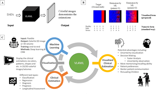
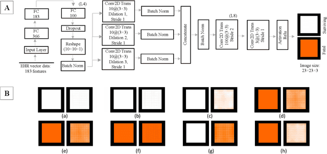
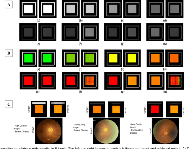
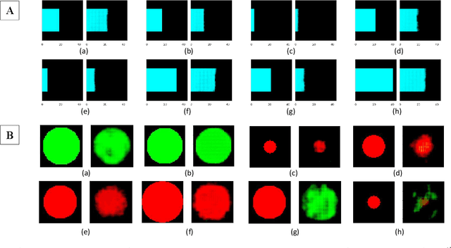
Abstract:With the advent of sophisticated machine learning (ML) techniques and the promising results they yield, especially in medical applications, where they have been investigated for different tasks to enhance the decision-making process. Since visualization is such an effective tool for human comprehension, memorization, and judgment, we have presented a first-of-its-kind estimation approach we refer to as Visualized Learning for Machine Learning (VL4ML) that not only can serve to assist physicians and clinicians in making reasoned medical decisions, but it also allows to appreciate the uncertainty visualization, which could raise incertitude in making the appropriate classification or prediction. For the proof of concept, and to demonstrate the generalized nature of this visualized estimation approach, five different case studies are examined for different types of tasks including classification, regression, and longitudinal prediction. A survey analysis with more than 100 individuals is also conducted to assess users' feedback on this visualized estimation method. The experiments and the survey demonstrate the practical merits of the VL4ML that include: (1) appreciating visually clinical/medical estimations; (2) getting closer to the patients' preferences; (3) improving doctor-patient communication, and (4) visualizing the uncertainty introduced through the black box effect of the deployed ML algorithm. All the source codes are shared via a GitHub repository.
Feasibility Assessment of Multitasking in MRI Neuroimaging Analysis: Tissue Segmentation, Cross-Modality Conversion and Bias correction
May 31, 2021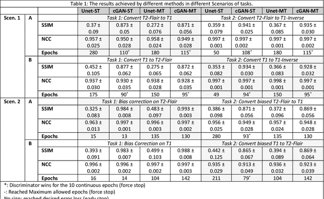
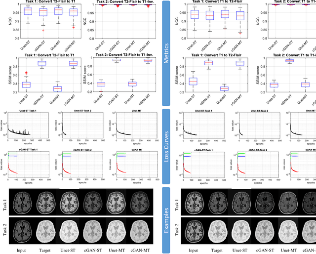
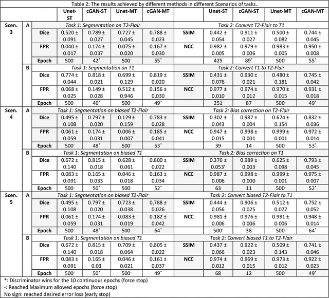
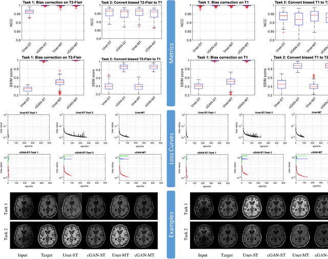
Abstract:Neuroimaging is essential in brain studies for the diagnosis and identification of disease, structure, and function of the brain in its healthy and disease states. Literature shows that there are advantages of multitasking with some deep learning (DL) schemes in challenging neuroimaging applications. This study examines the feasibility of using multitasking in three different applications, including tissue segmentation, cross-modality conversion, and bias-field correction. These applications reflect five different scenarios in which multitasking is explored and 280 training and testing sessions conducted for empirical evaluations. Two well-known networks, U-Net as a well-known convolutional neural network architecture, and a closed architecture based on the conditional generative adversarial network are implemented. Different metrics such as the normalized cross-correlation coefficient and Dice scores are used for comparison of methods and results of the different experiments. Statistical analysis is also provided by paired t-test. The present study explores the pros and cons of these methods and their practical impacts on multitasking in different implementation scenarios. This investigation shows that bias correction and cross-modality conversion applications are significantly easier than the segmentation application, and having multitasking with segmentation is not reasonable if one of them is identified as the main target application. However, when the main application is the segmentation of tissues, multitasking with cross-modality conversion is beneficial, especially for the U-net architecture.
SignCol: Open-Source Software for Collecting Sign Language Gestures
Oct 31, 2019


Abstract:Sign(ed) languages use gestures, such as hand or head movements, for communication. Sign language recognition is an assistive technology for individuals with hearing disability and its goal is to improve such individuals' life quality by facilitating their social involvement. Since sign languages are vastly varied in alphabets, as known as signs, a sign recognition software should be capable of handling eight different types of sign combinations, e.g. numbers, letters, words and sentences. Due to the intrinsic complexity and diversity of symbolic gestures, recognition algorithms need a comprehensive visual dataset to learn by. In this paper, we describe the design and implementation of a Microsoft Kinect-based open source software, called SignCol, for capturing and saving the gestures used in sign languages. Our work supports a multi-language database and reports the recorded items statistics. SignCol can capture and store colored(RGB) frames, depth frames, infrared frames, body index frames, coordinate mapped color-body frames, skeleton information of each frame and camera parameters simultaneously.
Image to Images Translation for Multi-Task Organ Segmentation and Bone Suppression in Chest X-Ray Radiography
Jun 24, 2019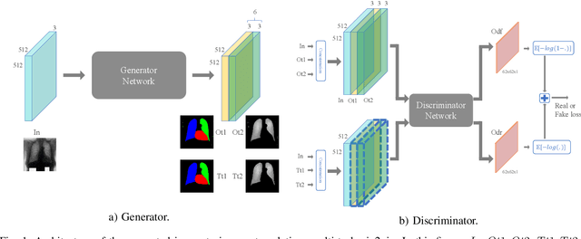


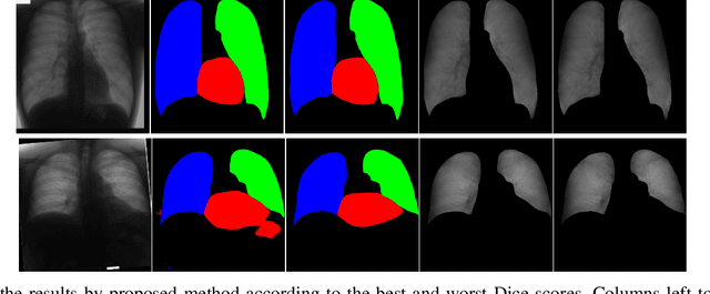
Abstract:Chest X-ray radiography is one of the earliest medical imaging technologies and remains one of the most widely-used for the diagnosis, screening and treatment follow up of diseases related to lungs and heart. The literature in this field of research reports many interesting studies dealing with the challenging tasks of bone suppression and organ segmentation but performed separately, limiting any learning that comes with the consolidation of parameters that could optimize both processes. Although image processing could facilitate computer aided diagnosis, machine learning seems more amenable in dealing with the many parameters one would have to contend with to yield an near optimal classification or decision-making process. This study, and for the first time, introduces a multitask deep learning model that generates simultaneously the bone-suppressed image and the organ segmented image, minimizing as a consequence the number of parameters the model has to deal with and optimizing the processing time as well; while at the same time exploiting the interplay in these parameters so as to benefit the performance of both tasks. The design architecture of this model, which relies on a conditional generative adversarial network, reveals the process on how we managed to modify the well-established pix2pix network to fit the need for multitasking and hence extending the standard image-to-image network to the new image-to-images architecture. Dilated convolutions are also used to improve the results through a more effective receptive field assessment. A comparison of the proposed approach to state-of-the-art algorithms is provided to gauge the merits of the proposed approach.
 Add to Chrome
 Add to Firefox
 Add to Edge