Ramtin Gharleghi
Machine-Learning Based Detection of Coronary Artery Calcification Using Synthetic Chest X-Rays
Nov 14, 2025Abstract:Coronary artery calcification (CAC) is a strong predictor of cardiovascular events, with CT-based Agatston scoring widely regarded as the clinical gold standard. However, CT is costly and impractical for large-scale screening, while chest X-rays (CXRs) are inexpensive but lack reliable ground truth labels, constraining deep learning development. Digitally reconstructed radiographs (DRRs) offer a scalable alternative by projecting CT volumes into CXR-like images while inheriting precise labels. In this work, we provide the first systematic evaluation of DRRs as a surrogate training domain for CAC detection. Using 667 CT scans from the COCA dataset, we generate synthetic DRRs and assess model capacity, super-resolution fidelity enhancement, preprocessing, and training strategies. Lightweight CNNs trained from scratch outperform large pretrained networks; pairing super-resolution with contrast enhancement yields significant gains; and curriculum learning stabilises training under weak supervision. Our best configuration achieves a mean AUC of 0.754, comparable to or exceeding prior CXR-based studies. These results establish DRRs as a scalable, label-rich foundation for CAC detection, while laying the foundation for future transfer learning and domain adaptation to real CXRs.
LWT-ARTERY-LABEL: A Lightweight Framework for Automated Coronary Artery Identification
Aug 09, 2025Abstract:Coronary artery disease (CAD) remains the leading cause of death globally, with computed tomography coronary angiography (CTCA) serving as a key diagnostic tool. However, coronary arterial analysis using CTCA, such as identifying artery-specific features from computational modelling, is labour-intensive and time-consuming. Automated anatomical labelling of coronary arteries offers a potential solution, yet the inherent anatomical variability of coronary trees presents a significant challenge. Traditional knowledge-based labelling methods fall short in leveraging data-driven insights, while recent deep-learning approaches often demand substantial computational resources and overlook critical clinical knowledge. To address these limitations, we propose a lightweight method that integrates anatomical knowledge with rule-based topology constraints for effective coronary artery labelling. Our approach achieves state-of-the-art performance on benchmark datasets, providing a promising alternative for automated coronary artery labelling.
Assessing Encoder-Decoder Architectures for Robust Coronary Artery Segmentation
Oct 16, 2023Abstract:Coronary artery diseases are among the leading causes of mortality worldwide. Timely and accurate diagnosis, facilitated by precise coronary artery segmentation, is pivotal in changing patient outcomes. In the realm of biomedical imaging, convolutional neural networks, especially the U-Net architecture, have revolutionised segmentation processes. However, one of the primary challenges remains the lack of benchmarking datasets specific to coronary arteries. However through the use of the recently published public dataset ASOCA, the potential of deep learning for accurate coronary segmentation can be improved. This paper delves deep into examining the performance of 25 distinct encoder-decoder combinations. Through analysis of the 40 cases provided to ASOCA participants, it is revealed that the EfficientNet-LinkNet combination, serving as encoder and decoder, stands out. It achieves a Dice coefficient of 0.882 and a 95th percentile Hausdorff distance of 4.753. These findings not only underscore the superiority of our model in comparison to those presented at the MICCAI 2020 challenge but also set the stage for future advancements in coronary artery segmentation, opening doors to enhanced diagnostic and treatment strategies.
MedShapeNet -- A Large-Scale Dataset of 3D Medical Shapes for Computer Vision
Sep 12, 2023


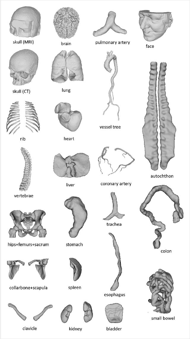
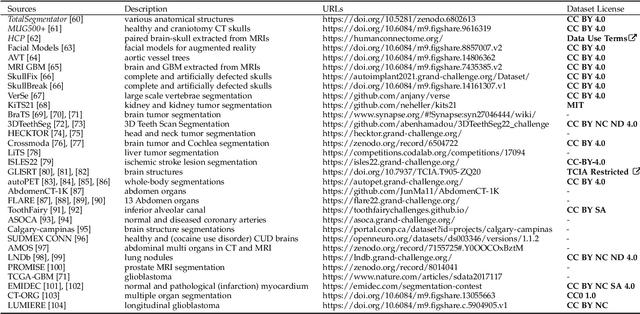
Abstract:We present MedShapeNet, a large collection of anatomical shapes (e.g., bones, organs, vessels) and 3D surgical instrument models. Prior to the deep learning era, the broad application of statistical shape models (SSMs) in medical image analysis is evidence that shapes have been commonly used to describe medical data. Nowadays, however, state-of-the-art (SOTA) deep learning algorithms in medical imaging are predominantly voxel-based. In computer vision, on the contrary, shapes (including, voxel occupancy grids, meshes, point clouds and implicit surface models) are preferred data representations in 3D, as seen from the numerous shape-related publications in premier vision conferences, such as the IEEE/CVF Conference on Computer Vision and Pattern Recognition (CVPR), as well as the increasing popularity of ShapeNet (about 51,300 models) and Princeton ModelNet (127,915 models) in computer vision research. MedShapeNet is created as an alternative to these commonly used shape benchmarks to facilitate the translation of data-driven vision algorithms to medical applications, and it extends the opportunities to adapt SOTA vision algorithms to solve critical medical problems. Besides, the majority of the medical shapes in MedShapeNet are modeled directly on the imaging data of real patients, and therefore it complements well existing shape benchmarks comprising of computer-aided design (CAD) models. MedShapeNet currently includes more than 100,000 medical shapes, and provides annotations in the form of paired data. It is therefore also a freely available repository of 3D models for extended reality (virtual reality - VR, augmented reality - AR, mixed reality - MR) and medical 3D printing. This white paper describes in detail the motivations behind MedShapeNet, the shape acquisition procedures, the use cases, as well as the usage of the online shape search portal: https://medshapenet.ikim.nrw/
Computed tomography coronary angiogram images, annotations and associated data of normal and diseased arteries
Nov 03, 2022Abstract:Computed Tomography Coronary Angiography (CTCA) is a non-invasive method to evaluate coronary artery anatomy and disease. CTCA is ideal for geometry reconstruction to create virtual models of coronary arteries. To our knowledge there is no public dataset that includes centrelines and segmentation of the full coronary tree. We provide anonymized CTCA images, voxel-wise annotations and associated data in the form of centrelines, calcification scores and meshes of the coronary lumen in 20 normal and 20 diseased cases. Images were obtained along with patient information with informed, written consent as part of Coronary Atlas (https://www.coronaryatlas.org/). Cases were classified as normal (zero calcium score with no signs of stenosis) or diseased (confirmed coronary artery disease). Manual voxel-wise segmentations by three experts were combined using majority voting to generate the final annotations. Provided data can be used for a variety of research purposes, such as 3D printing patient-specific models, development and validation of segmentation algorithms, education and training of medical personnel and in-silico analyses such as testing of medical devices.
 Add to Chrome
 Add to Firefox
 Add to Edge