Michael K. Atalay
Abn-BLIP: Abnormality-aligned Bootstrapping Language-Image Pre-training for Pulmonary Embolism Diagnosis and Report Generation from CTPA
Mar 03, 2025Abstract:Medical imaging plays a pivotal role in modern healthcare, with computed tomography pulmonary angiography (CTPA) being a critical tool for diagnosing pulmonary embolism and other thoracic conditions. However, the complexity of interpreting CTPA scans and generating accurate radiology reports remains a significant challenge. This paper introduces Abn-BLIP (Abnormality-aligned Bootstrapping Language-Image Pretraining), an advanced diagnosis model designed to align abnormal findings to generate the accuracy and comprehensiveness of radiology reports. By leveraging learnable queries and cross-modal attention mechanisms, our model demonstrates superior performance in detecting abnormalities, reducing missed findings, and generating structured reports compared to existing methods. Our experiments show that Abn-BLIP outperforms state-of-the-art medical vision-language models and 3D report generation methods in both accuracy and clinical relevance. These results highlight the potential of integrating multimodal learning strategies for improving radiology reporting. The source code is available at https://github.com/zzs95/abn-blip.
Pulmonary Embolism Mortality Prediction Using Multimodal Learning Based on Computed Tomography Angiography and Clinical Data
Jun 03, 2024Abstract:Purpose: Pulmonary embolism (PE) is a significant cause of mortality in the United States. The objective of this study is to implement deep learning (DL) models using Computed Tomography Pulmonary Angiography (CTPA), clinical data, and PE Severity Index (PESI) scores to predict PE mortality. Materials and Methods: 918 patients (median age 64 years, range 13-99 years, 52% female) with 3,978 CTPAs were identified via retrospective review across three institutions. To predict survival, an AI model was used to extract disease-related imaging features from CTPAs. Imaging features and/or clinical variables were then incorporated into DL models to predict survival outcomes. Four models were developed as follows: (1) using CTPA imaging features only; (2) using clinical variables only; (3) multimodal, integrating both CTPA and clinical variables; and (4) multimodal fused with calculated PESI score. Performance and contribution from each modality were evaluated using concordance index (c-index) and Net Reclassification Improvement, respectively. Performance was compared to PESI predictions using the Wilcoxon signed-rank test. Kaplan-Meier analysis was performed to stratify patients into high- and low-risk groups. Additional factor-risk analysis was conducted to account for right ventricular (RV) dysfunction. Results: For both data sets, the PESI-fused and multimodal models achieved higher c-indices than PESI alone. Following stratification of patients into high- and low-risk groups by multimodal and PESI-fused models, mortality outcomes differed significantly (both p<0.001). A strong correlation was found between high-risk grouping and RV dysfunction. Conclusions: Multiomic DL models incorporating CTPA features, clinical data, and PESI achieved higher c-indices than PESI alone for PE survival prediction.
Region-specific Risk Quantification for Interpretable Prognosis of COVID-19
May 05, 2024


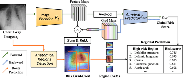
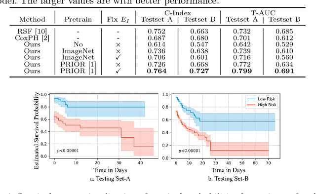
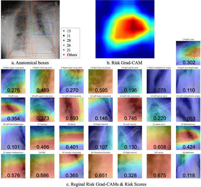
Abstract:The COVID-19 pandemic has strained global public health, necessitating accurate diagnosis and intervention to control disease spread and reduce mortality rates. This paper introduces an interpretable deep survival prediction model designed specifically for improved understanding and trust in COVID-19 prognosis using chest X-ray (CXR) images. By integrating a large-scale pretrained image encoder, Risk-specific Grad-CAM, and anatomical region detection techniques, our approach produces regional interpretable outcomes that effectively capture essential disease features while focusing on rare but critical abnormal regions. Our model's predictive results provide enhanced clarity and transparency through risk area localization, enabling clinicians to make informed decisions regarding COVID-19 diagnosis with better understanding of prognostic insights. We evaluate the proposed method on a multi-center survival dataset and demonstrate its effectiveness via quantitative and qualitative assessments, achieving superior C-indexes (0.764 and 0.727) and time-dependent AUCs (0.799 and 0.691). These results suggest that our explainable deep survival prediction model surpasses traditional survival analysis methods in risk prediction, improving interpretability for clinical decision making and enhancing AI system trustworthiness.
 Add to Chrome
 Add to Firefox
 Add to Edge