Hamid Moradi
Automatic Search for Photoacoustic Marker Using Automated Transrectal Ultrasound
Jul 20, 2023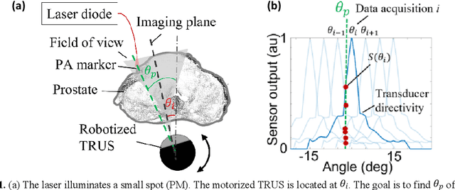

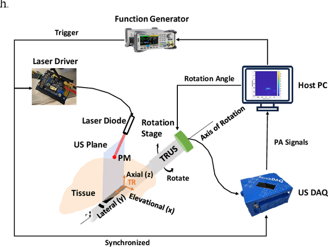
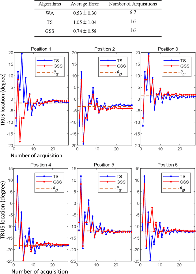
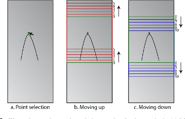
Abstract:Real-time transrectal ultrasound (TRUS) image guidance during robot-assisted laparoscopic radical prostatectomy has the potential to enhance surgery outcomes. Whether conventional or photoacoustic TRUS is used, the robotic system and the TRUS must be registered to each other. Accurate registration can be performed using photoacoustic (PA markers). However, this requires a manual search by an assistant [19]. This paper introduces the first automatic search for PA markers using a transrectal ultrasound robot. This effectively reduces the challenges associated with the da Vinci-TRUS registration. This paper investigated the performance of three search algorithms in simulation and experiment: Weighted Average (WA), Golden Section Search (GSS), and Ternary Search (TS). For validation, a surgical prostate scenario was mimicked and various ex vivo tissues were tested. As a result, the WA algorithm can achieve 0.53 degree average error after 9 data acquisitions, while the TS and GSS algorithm can achieve 0.29 degree and 0.48 degree average errors after 28 data acquisitions.
Arc-to-line frame registration method for ultrasound and photoacoustic image-guided intraoperative robot-assisted laparoscopic prostatectomy
Jun 21, 2023


Abstract:Purpose: To achieve effective robot-assisted laparoscopic prostatectomy, the integration of transrectal ultrasound (TRUS) imaging system which is the most widely used imaging modelity in prostate imaging is essential. However, manual manipulation of the ultrasound transducer during the procedure will significantly interfere with the surgery. Therefore, we propose an image co-registration algorithm based on a photoacoustic marker method, where the ultrasound / photoacoustic (US/PA) images can be registered to the endoscopic camera images to ultimately enable the TRUS transducer to automatically track the surgical instrument Methods: An optimization-based algorithm is proposed to co-register the images from the two different imaging modalities. The principles of light propagation and an uncertainty in PM detection were assumed in this algorithm to improve the stability and accuracy of the algorithm. The algorithm is validated using the previously developed US/PA image-guided system with a da Vinci surgical robot. Results: The target-registration-error (TRE) is measured to evaluate the proposed algorithm. In both simulation and experimental demonstration, the proposed algorithm achieved a sub-centimeter accuracy which is acceptable in practical clinics. The result is also comparable with our previous approach, and the proposed method can be implemented with a normal white light stereo camera and doesn't require highly accurate localization of the PM. Conclusion: The proposed frame registration algorithm enabled a simple yet efficient integration of commercial US/PA imaging system into laparoscopic surgical setting by leveraging the characteristic properties of acoustic wave propagation and laser excitation, contributing to automated US/PA image-guided surgical intervention applications.
Single Frame Laser Diode Photoacoustic Imaging: Denoising and Reconstruction
Dec 16, 2022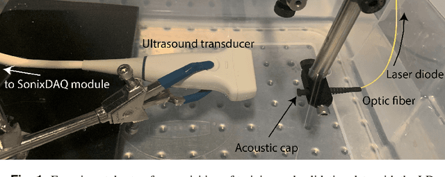
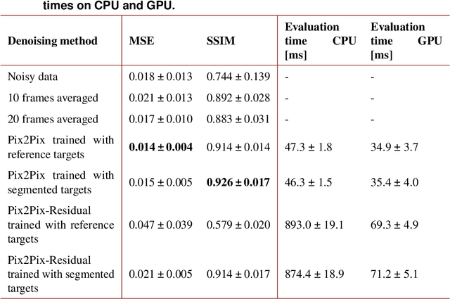
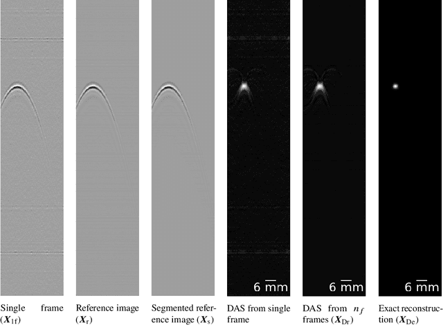
Abstract:A new development in photoacoustic (PA) imaging has been the use of compact, portable and low-cost laser diodes (LDs), but LD-based PA imaging suffers from low signal intensity recorded by the conventional transducers. A common method to improve signal strength is temporal averaging, which reduces frame rate and increases laser exposure to patients. To tackle this problem, we propose a deep learning method that will denoise the PA images before beamforming with a very few frames, even one. We also present a deep learning method to automatically reconstruct point sources from noisy pre-beamformed data. Finally, we employ a strategy of combined denoising and reconstruction, which can supplement the reconstruction algorithm for very low signal-to-noise ratio inputs.
Quasi-Real Time Multi-Frequency 3D Shear Wave Absolute Vibro-Elastography System for Prostate
May 09, 2022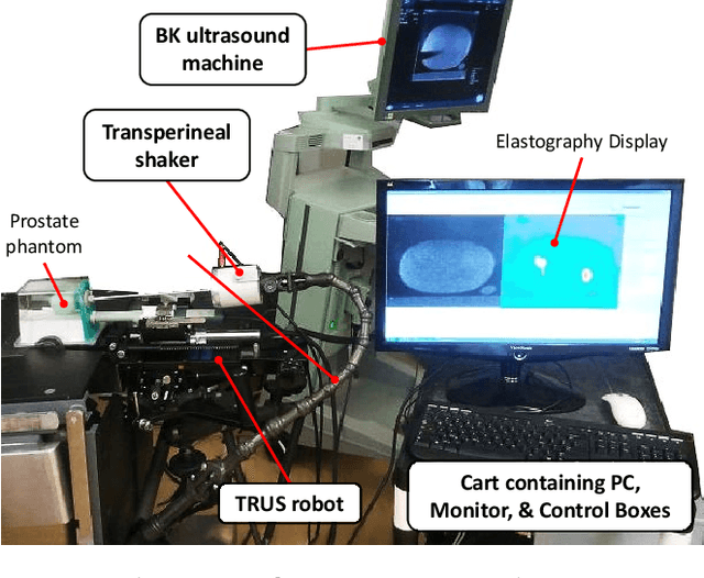
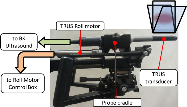
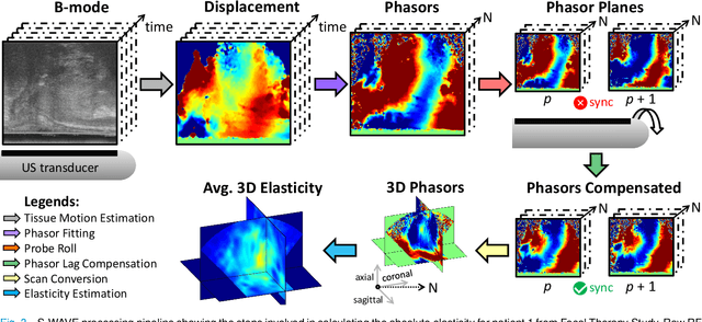
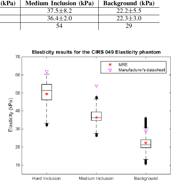
Abstract:This article describes a novel quasi-real time system for quantitative and volumetric measurement of tissue elasticity in the prostate. Tissue elasticity is computed by using a local frequency estimator to measure the three dimensional local wavelengths of a steady-state shear wave within the prostate gland. The shear wave is created using a mechanical voice coil shaker which transmits multi-frequency vibrations transperineally. Radio frequency data is streamed directly from a BK Medical 8848 trans-rectal ultrasound transducer to an external computer where tissue displacement due to the excitation is measured using a speckle tracking algorithm. Bandpass sampling is used that eliminates the need for an ultra fast frame rate to track the tissue motion and allows for accurate reconstruction at a sampling frequency that is below the Nyquist rate. A roll motor with computer control is used to rotate the sagittal array of the transducer and obtain the 3D data. Two CIRS phantoms were used to validate both the accuracy of the elasticity measurement as well as the functional feasibility of using the system for in vivo prostate imaging. The system has been used in two separate clinical studies as a method for cancer identification. The results, presented here, show numerical and visual correlations between our stiffness measurements and cancer likelihood as determined from pathology results. Initial published results using this system include an area under the receiver operating characteristic curve of 0.82+/-0.01 with regards to prostate cancer identification in the peripheral zone.
 Add to Chrome
 Add to Firefox
 Add to Edge