Quasi-Real Time Multi-Frequency 3D Shear Wave Absolute Vibro-Elastography System for Prostate
Paper and Code
May 09, 2022


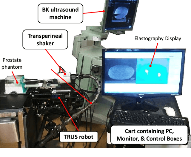
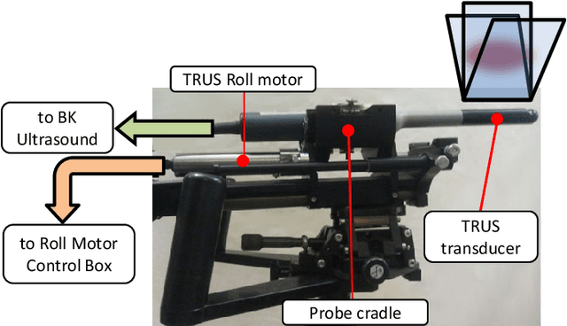
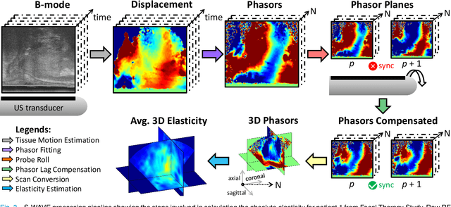
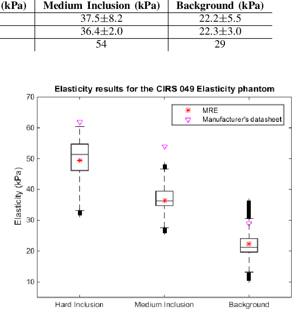
This article describes a novel quasi-real time system for quantitative and volumetric measurement of tissue elasticity in the prostate. Tissue elasticity is computed by using a local frequency estimator to measure the three dimensional local wavelengths of a steady-state shear wave within the prostate gland. The shear wave is created using a mechanical voice coil shaker which transmits multi-frequency vibrations transperineally. Radio frequency data is streamed directly from a BK Medical 8848 trans-rectal ultrasound transducer to an external computer where tissue displacement due to the excitation is measured using a speckle tracking algorithm. Bandpass sampling is used that eliminates the need for an ultra fast frame rate to track the tissue motion and allows for accurate reconstruction at a sampling frequency that is below the Nyquist rate. A roll motor with computer control is used to rotate the sagittal array of the transducer and obtain the 3D data. Two CIRS phantoms were used to validate both the accuracy of the elasticity measurement as well as the functional feasibility of using the system for in vivo prostate imaging. The system has been used in two separate clinical studies as a method for cancer identification. The results, presented here, show numerical and visual correlations between our stiffness measurements and cancer likelihood as determined from pathology results. Initial published results using this system include an area under the receiver operating characteristic curve of 0.82+/-0.01 with regards to prostate cancer identification in the peripheral zone.
 Add to Chrome
 Add to Firefox
 Add to Edge