Anjali Agrawal
An Introduction to Reinforcement Learning: Fundamental Concepts and Practical Applications
Aug 13, 2024Abstract:Reinforcement Learning (RL) is a branch of Artificial Intelligence (AI) which focuses on training agents to make decisions by interacting with their environment to maximize cumulative rewards. An overview of RL is provided in this paper, which discusses its core concepts, methodologies, recent trends, and resources for learning. We provide a detailed explanation of key components of RL such as states, actions, policies, and reward signals so that the reader can build a foundational understanding. The paper also provides examples of various RL algorithms, including model-free and model-based methods. In addition, RL algorithms are introduced and resources for learning and implementing them are provided, such as books, courses, and online communities. This paper demystifies a comprehensive yet simple introduction for beginners by offering a structured and clear pathway for acquiring and implementing real-time techniques.
Deep Learning for Automated Screening of Tuberculosis from Indian Chest X-rays: Analysis and Update
Nov 19, 2020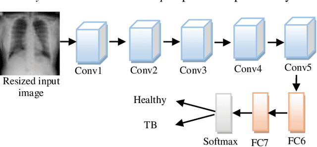
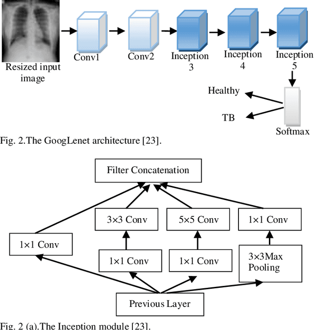
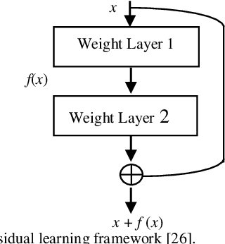
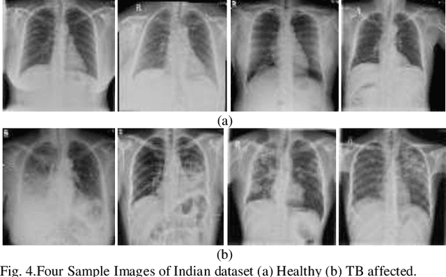
Abstract:Background and Objective: Tuberculosis (TB) is a significant public health issue and a leading cause of death worldwide. Millions of deaths can be averted by early diagnosis and successful treatment of TB patients. Automated diagnosis of TB holds vast potential to assist medical experts in expediting and improving its diagnosis, especially in developing countries like India, where there is a shortage of trained medical experts and radiologists. To date, several deep learning based methods for automated detection of TB from chest radiographs have been proposed. However, the performance of a few of these methods on the Indian chest radiograph data set has been suboptimal, possibly due to different texture of the lungs on chest radiographs of Indian subjects compared to other countries. Thus deep learning for accurate and automated diagnosis of TB on Indian datasets remains an important subject of research. Methods: The proposed work explores the performance of convolutional neural networks (CNNs) for the diagnosis of TB in Indian chest x-ray images. Three different pre-trained neural network models, AlexNet, GoogLenet, and ResNet are used to classify chest x-ray images into healthy or TB infected. The proposed approach does not require any pre-processing technique. Also, other works use pre-trained NNs as a tool for crafting features and then apply standard classification techniques. However, we attempt an end to end NN model based diagnosis of TB from chest x-rays. The proposed visualization tool can also be used by radiologists in the screening of large datasets. Results: The proposed method achieved 93.40% accuracy with 98.60% sensitivity to diagnose TB for the Indian population. Conclusions: The performance of the proposed method is also tested against techniques described in the literature. The proposed method outperforms the state of art on Indian and Shenzhen datasets.
Deep LF-Net: Semantic Lung Segmentation from Indian Chest Radiographs Including Severely Unhealthy Images
Nov 19, 2020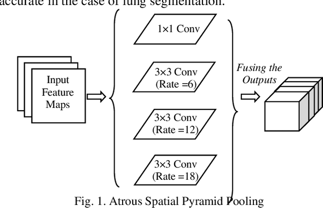
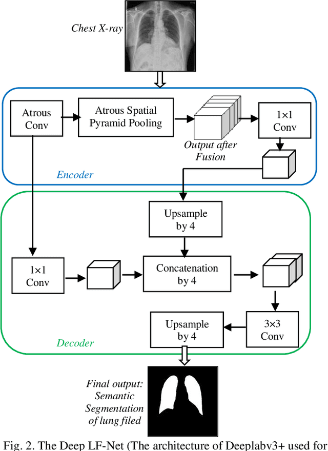
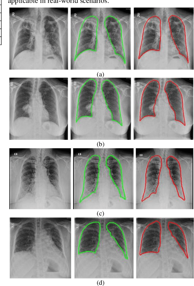
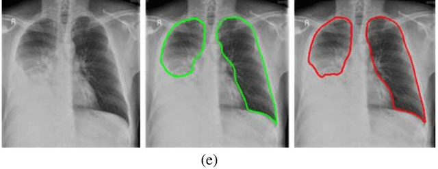
Abstract:A chest radiograph, commonly called chest x-ray (CxR), plays a vital role in the diagnosis of various lung diseases, such as lung cancer, tuberculosis, pneumonia, and many more. Automated segmentation of the lungs is an important step to design a computer-aided diagnostic tool for examination of a CxR. Precise lung segmentation is considered extremely challenging because of variance in the shape of the lung caused by health issues, age, and gender. The proposed work investigates the use of an efficient deep convolutional neural network for accurate segmentation of lungs from CxR. We attempt an end to end DeepLabv3+ network which integrates DeepLab architecture, encoder-decoder, and dilated convolution for semantic lung segmentation with fast training and high accuracy. We experimented with the different pre-trained base networks: Resnet18 and Mobilenetv2, associated with the Deeplabv3+ model for performance analysis. The proposed approach does not require any pre-processing technique on chest x-ray images before being fed to a neural network. Morphological operations were used to remove false positives that occurred during semantic segmentation. We construct a CxR dataset of the Indian population that contain healthy and unhealthy CxRs of clinically confirmed patients of tuberculosis, chronic obstructive pulmonary disease, interstitial lung disease, pleural effusion, and lung cancer. The proposed method is tested on 688 images of our Indian CxR dataset including images with severe abnormal findings to validate its robustness. We also experimented on commonly used benchmark datasets such as Japanese Society of Radiological Technology; Montgomery County, USA; and Shenzhen, China for state-of-the-art comparison. The performance of our method is tested against techniques described in the literature and achieved the highest accuracy for lung segmentation on Indian and public datasets.
Multi-Task Driven Explainable Diagnosis of COVID-19 using Chest X-ray Images
Aug 03, 2020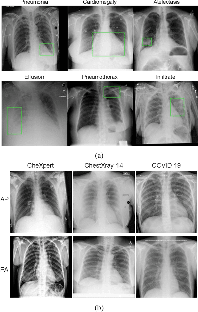
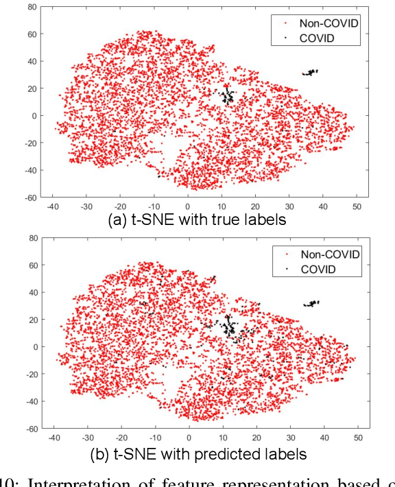
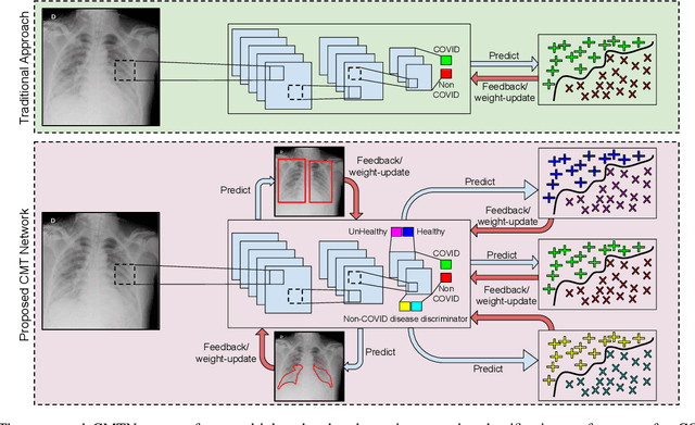
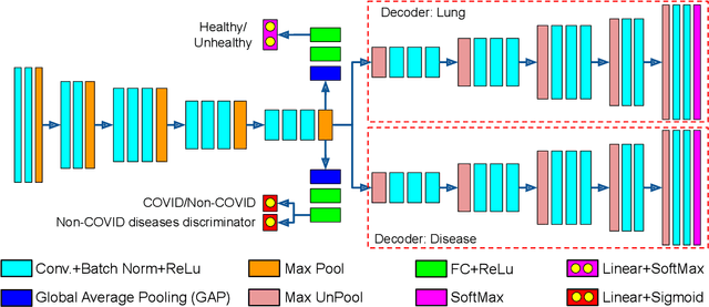
Abstract:With increasing number of COVID-19 cases globally, all the countries are ramping up the testing numbers. While the RT-PCR kits are available in sufficient quantity in several countries, others are facing challenges with limited availability of testing kits and processing centers in remote areas. This has motivated researchers to find alternate methods of testing which are reliable, easily accessible and faster. Chest X-Ray is one of the modalities that is gaining acceptance as a screening modality. Towards this direction, the paper has two primary contributions. Firstly, we present the COVID-19 Multi-Task Network which is an automated end-to-end network for COVID-19 screening. The proposed network not only predicts whether the CXR has COVID-19 features present or not, it also performs semantic segmentation of the regions of interest to make the model explainable. Secondly, with the help of medical professionals, we manually annotate the lung regions of 9000 frontal chest radiographs taken from ChestXray-14, CheXpert and a consolidated COVID-19 dataset. Further, 200 chest radiographs pertaining to COVID-19 patients are also annotated for semantic segmentation. This database will be released to the research community.
 Add to Chrome
 Add to Firefox
 Add to Edge