Zichao Bian
Ptychographic sensor for large-scale lensless microbial monitoring with high spatiotemporal resolution
Dec 15, 2021
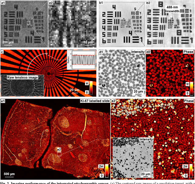

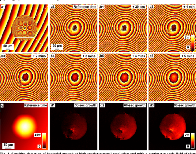
Abstract:Traditional microbial detection methods often rely on the overall property of microbial cultures and cannot resolve individual growth event at high spatiotemporal resolution. As a result, they require bacteria to grow to confluence and then interpret the results. Here, we demonstrate the application of an integrated ptychographic sensor for lensless cytometric analysis of microbial cultures over a large scale and with high spatiotemporal resolution. The reported device can be placed within a regular incubator or used as a standalone incubating unit for long-term microbial monitoring. For longitudinal study where massive data are acquired at sequential time points, we report a new temporal-similarity constraint to increase the temporal resolution of ptychographic reconstruction by 7-fold. With this strategy, the reported device achieves a centimeter-scale field of view, a half-pitch spatial resolution of 488 nm, and a temporal resolution of 15-second intervals. For the first time, we report the direct observation of bacterial growth in a 15-second interval by tracking the phase wraps of the recovered images, with high phase sensitivity like that in interferometric measurements. We also characterize cell growth via longitudinal dry mass measurement and perform rapid bacterial detection at low concentrations. For drug-screening application, we demonstrate proof-of-concept antibiotic susceptibility testing and perform single-cell analysis of antibiotic-induced filamentation. The combination of high phase sensitivity, high spatiotemporal resolution, and large field of view is unique among existing microscopy techniques. As a quantitative and miniaturized platform, it can improve studies with microorganisms and other biospecimens at resource-limited settings.
High-throughput lensless whole slide imaging via continuous height-varying modulation of tilted sensor
Sep 28, 2021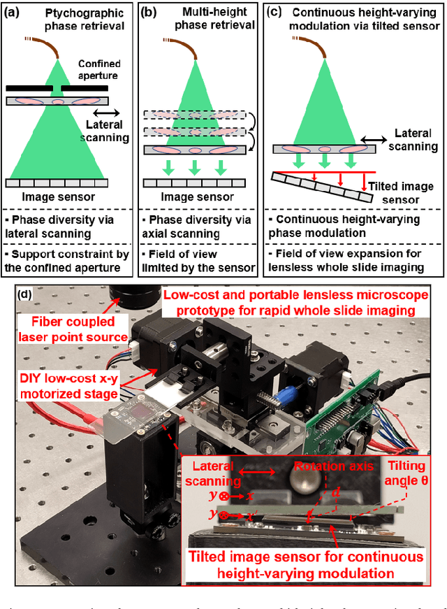
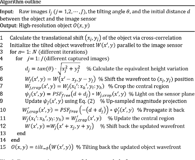

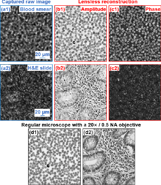
Abstract:We report a new lensless microscopy configuration by integrating the concepts of transverse translational ptychography and defocus multi-height phase retrieval. In this approach, we place a tilted image sensor under the specimen for linearly-increasing phase modulation along one lateral direction. Similar to the operation of ptychography, we laterally translate the specimen and acquire the diffraction images for reconstruction. Since the axial distance between the specimen and the sensor varies at different lateral positions, laterally translating the specimen effectively introduces defocus multi-height measurements while eliminating axial scanning. Lateral translation further introduces sub-pixel shift for pixel super-resolution imaging and naturally expands the field of view for rapid whole slide imaging. We show that the equivalent height variation can be precisely estimated from the lateral shift of the specimen, thereby addressing the challenge of precise axial positioning in conventional multi-height phase retrieval. Using a sensor with a 1.67-micron pixel size, our low-cost and field-portable prototype can resolve 690-nm linewidth on the resolution target. We show that a whole slide image of a blood smear with a 120-mm^2 field of view can be acquired in 18 seconds. We also demonstrate accurate automatic white blood cell counting from the recovered image. The reported approach may provide a turnkey solution for addressing point-of-care- and telemedicine-related challenges.
Bypassing the resolution limit of diffractive zone plate optics via rotational Fourier ptychography
Feb 07, 2021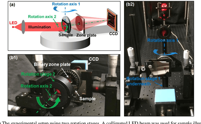
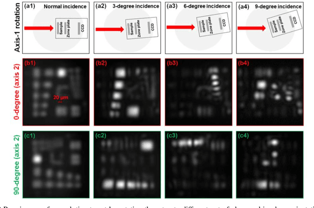
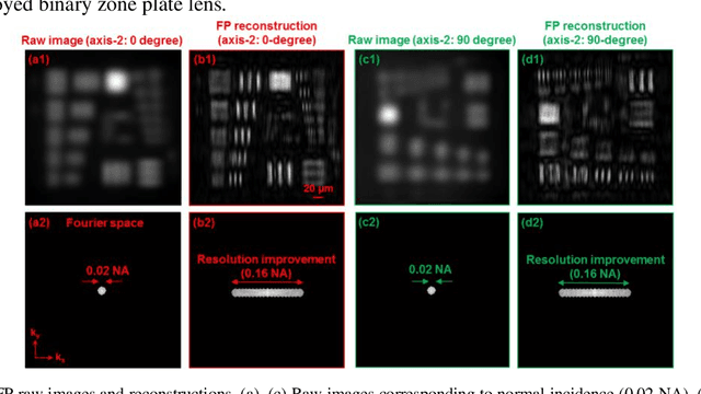
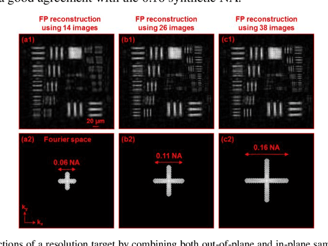
Abstract:Diffractive zone plate optics uses a thin micro-structure pattern to alter the propagation direction of the incoming light wave. It has found important applications in extreme-wavelength imaging where conventional refractive lenses do not exist. The resolution limit of zone plate optics is determined by the smallest width of the outermost zone. In order to improve the achievable resolution, significant efforts have been devoted to the fabrication of very small zone width with ultrahigh placement accuracy. Here, we report the use of a diffractometer setup for bypassing the resolution limit of zone plate optics. In our prototype, we mounted the sample on two rotation stages and used a low-resolution binary zone plate to relay the sample plane to the detector. We then performed both in-plane and out-of-plane sample rotations and captured the corresponding raw images. The captured images were processed using a Fourier ptychographic procedure for resolution improvement. The final achievable resolution of the reported setup is not determined by the smallest width structures of the employed binary zone plate; instead, it is determined by the maximum angle of the out-of-plane rotation. In our experiment, we demonstrated 8-fold resolution improvement using both a resolution target and a titanium dioxide sample. The reported approach may be able to bypass the fabrication challenge of diffractive elements and open up new avenues for microscopy with extreme wavelengths.
Axially-shifted pattern illumination for macroscale turbidity suppression and virtual volumetric confocal imaging without axial scanning
Dec 14, 2018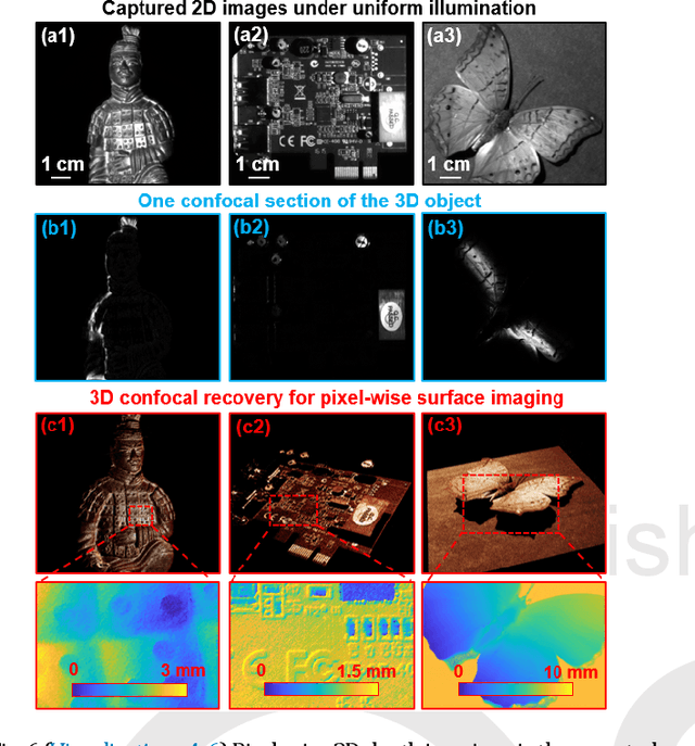
Abstract:Structured illumination has been widely used for optical sectioning and 3D surface recovery. In a typical implementation, multiple images under non-uniform pattern illumination are used to recover a single object section. Axial scanning of the sample or the objective lens is needed for acquiring the 3D volumetric data. Here we demonstrate the use of axially-shifted pattern illumination (asPI) for virtual volumetric confocal imaging without axial scanning. In the reported approach, we project illumination patterns at a tilted angle with respect to the detection optics. As such, the illumination patterns shift laterally at different z sections and the sample information at different z-sections can be recovered based on the captured 2D images. We demonstrate the reported approach for virtual confocal imaging through a diffusing layer and underwater 3D imaging through diluted milk. We show that we can acquire the entire confocal volume in ~1s with a throughput of 420 megapixels per second. Our approach may provide new insights for developing confocal light ranging and detection systems in degraded visual environments.
Rapid focus map surveying for whole slide imaging with continues sample motion
Jul 06, 2017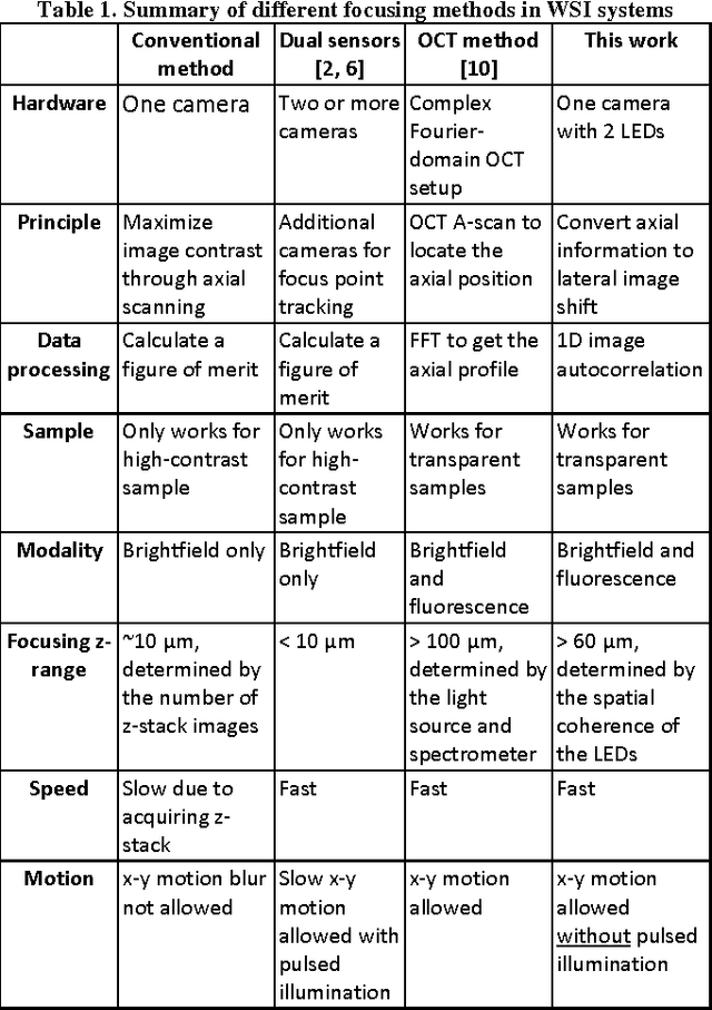
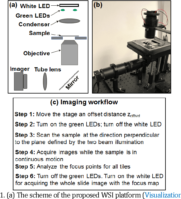
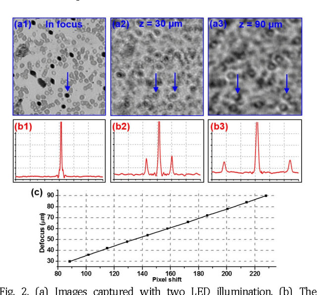
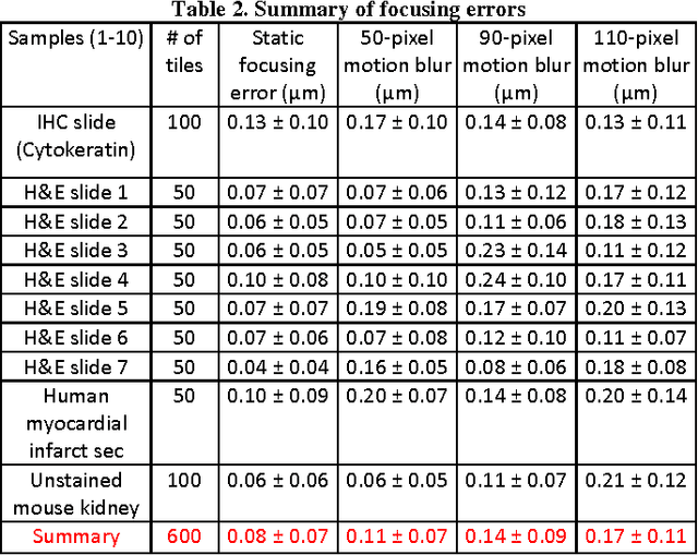
Abstract:Whole slide imaging (WSI) has recently been cleared for primary diagnosis in the US. A critical challenge of WSI is to perform accurate focusing in high speed. Traditional systems create a focus map prior to scanning. For each focus point on the map, sample needs to be static in the x-y plane and axial scanning is needed to maximize the contrast. Here we report a novel focus map surveying method for WSI. The reported method requires no axial scanning, no additional camera and lens, works for stained and transparent samples, and allows continuous sample motion in the surveying process. It can be used for both brightfield and fluorescence WSI. By using a 20X, 0.75 NA objective lens, we demonstrate a mean focusing error of ~0.08 microns in the static mode and ~0.17 microns in the continuous motion mode. The reported method may provide a turnkey solution for most existing WSI systems for its simplicity, robustness, accuracy, and high-speed. It may also standardize the imaging performance of WSI systems for digital pathology and find other applications in high-content microscopy such as DNA sequencing and time-lapse live-cell imaging.
 Add to Chrome
 Add to Firefox
 Add to Edge