Xiaopeng Shao
Reconstructing Images of Two Adjacent Objects through Scattering Medium Using Generative Adversarial Network
Jul 24, 2021


Abstract:Reconstruction of image by using convolutional neural networks (CNNs) has been vigorously studied in the last decade. Until now, there have being developed several techniques for imaging of a single object through scattering medium by using neural networks, however how to reconstruct images of more than one object simultaneously seems hard to realize. In this paper, we demonstrate an approach by using generative adversarial network (GAN) to reconstruct images of two adjacent objects through scattering media. We construct an imaging system for imaging of two adjacent objects behind the scattering media. In general, as the light field of two adjacent object images pass through the scattering slab, a speckle pattern is obtained. The designed adversarial network, which is called as YGAN, is employed to reconstruct the images simultaneously. It is shown that based on the trained YGAN, we can reconstruct images of two adjacent objects from one speckle pattern with high fidelity. In addition, we study the influence of the object image types, and the distance between the two adjacent objects on the fidelity of the reconstructed images. Moreover even if another scattering medium is inserted between the two objects, we can also reconstruct the images of two objects from a speckle with high quality. The technique presented in this work can be used for applications in areas of medical image analysis, such as medical image classification, segmentation, and studies of multi-object scattering imaging etc.
Large field-of-view non-invasive imaging through scattering layers using fluctuating random illumination
Jul 17, 2021


Abstract:On-invasive optical imaging techniques are essential diagnostic tools in many fields. Although various recent methods have been proposed to utilize and control light in multiple scattering media, non-invasive optical imaging through and inside scattering layers across a large field of view remains elusive due to the physical limits set by the optical memory effect, especially without wavefront shaping techniques. Here, we demonstrate an approach that enables non-invasive fluorescence imaging behind scattering layers with field-of-views extending well beyond the optical memory effect. The method consists in demixing the speckle patterns emitted by a fluorescent object under variable unknown random illumination, using matrix factorization and a novel fingerprint-based reconstruction. Experimental validation shows the efficiency and robustness of the method with various fluorescent samples, covering a field of view up to three times the optical memory effect range. Our non-invasive imaging technique is simple, neither requires a spatial light modulator nor a guide star, and can be generalized to a wide range of incoherent contrast mechanisms and illumination schemes.
Bypassing the resolution limit of diffractive zone plate optics via rotational Fourier ptychography
Feb 07, 2021


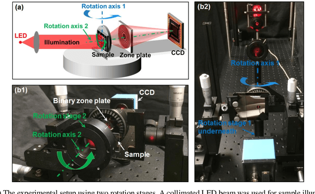
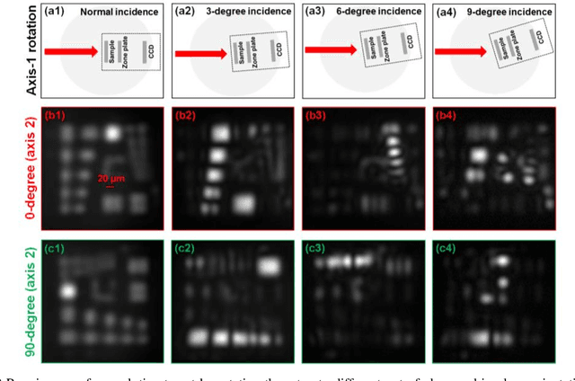
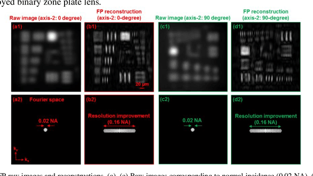
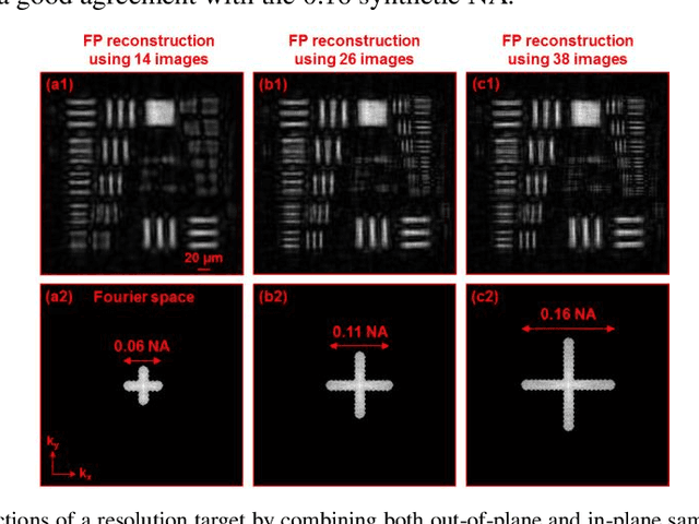
Abstract:Diffractive zone plate optics uses a thin micro-structure pattern to alter the propagation direction of the incoming light wave. It has found important applications in extreme-wavelength imaging where conventional refractive lenses do not exist. The resolution limit of zone plate optics is determined by the smallest width of the outermost zone. In order to improve the achievable resolution, significant efforts have been devoted to the fabrication of very small zone width with ultrahigh placement accuracy. Here, we report the use of a diffractometer setup for bypassing the resolution limit of zone plate optics. In our prototype, we mounted the sample on two rotation stages and used a low-resolution binary zone plate to relay the sample plane to the detector. We then performed both in-plane and out-of-plane sample rotations and captured the corresponding raw images. The captured images were processed using a Fourier ptychographic procedure for resolution improvement. The final achievable resolution of the reported setup is not determined by the smallest width structures of the employed binary zone plate; instead, it is determined by the maximum angle of the out-of-plane rotation. In our experiment, we demonstrated 8-fold resolution improvement using both a resolution target and a titanium dioxide sample. The reported approach may be able to bypass the fabrication challenge of diffractive elements and open up new avenues for microscopy with extreme wavelengths.
 Add to Chrome
 Add to Firefox
 Add to Edge