Kaikai Guo
Solving Fourier ptychographic imaging problems via neural network modeling and TensorFlow
Mar 09, 2018

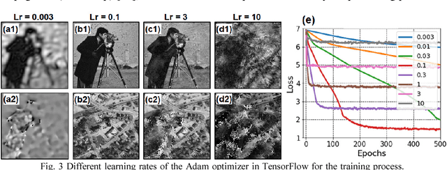
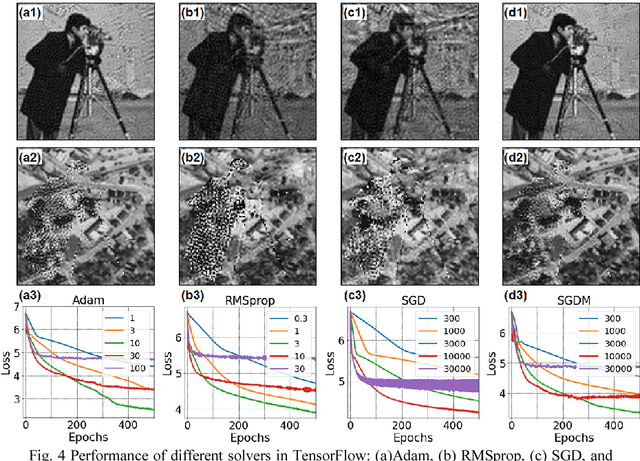
Abstract:Fourier ptychography is a recently developed imaging approach for large field-of-view and high-resolution microscopy. Here we model the Fourier ptychographic forward imaging process using a convolution neural network (CNN) and recover the complex object information in the network training process. In this approach, the input of the network is the point spread function in the spatial domain or the coherent transfer function in the Fourier domain. The object is treated as 2D learnable weights of a convolution or a multiplication layer. The output of the network is modeled as the loss function we aim to minimize. The batch size of the network corresponds to the number of captured low-resolution images in one forward / backward pass. We use a popular open-source machine learning library, TensorFlow, for setting up the network and conducting the optimization process. We analyze the performance of different learning rates, different solvers, and different batch sizes. It is shown that a large batch size with the Adam optimizer achieves the best performance in general. To accelerate the phase retrieval process, we also discuss a strategy to implement Fourier-magnitude projection using a multiplication neural network model. Since convolution and multiplication are the two most-common operations in imaging modeling, the reported approach may provide a new perspective to examine many coherent and incoherent systems. As a demonstration, we discuss the extensions of the reported networks for modeling single-pixel imaging and structured illumination microscopy (SIM). 4-frame resolution doubling is demonstrated using a neural network for SIM. We have made our implementation code open-source for the broad research community.
Rapid focus map surveying for whole slide imaging with continues sample motion
Jul 06, 2017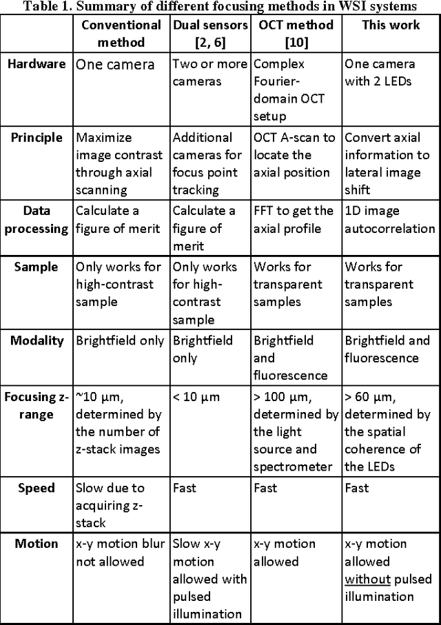
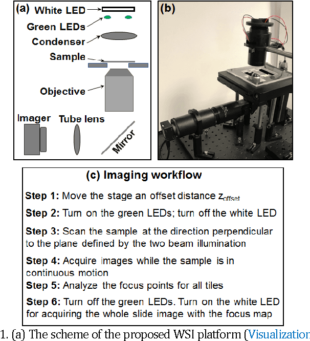
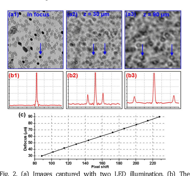
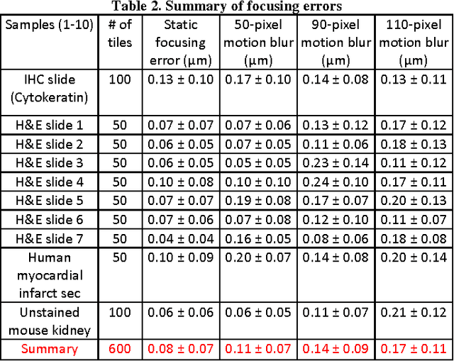
Abstract:Whole slide imaging (WSI) has recently been cleared for primary diagnosis in the US. A critical challenge of WSI is to perform accurate focusing in high speed. Traditional systems create a focus map prior to scanning. For each focus point on the map, sample needs to be static in the x-y plane and axial scanning is needed to maximize the contrast. Here we report a novel focus map surveying method for WSI. The reported method requires no axial scanning, no additional camera and lens, works for stained and transparent samples, and allows continuous sample motion in the surveying process. It can be used for both brightfield and fluorescence WSI. By using a 20X, 0.75 NA objective lens, we demonstrate a mean focusing error of ~0.08 microns in the static mode and ~0.17 microns in the continuous motion mode. The reported method may provide a turnkey solution for most existing WSI systems for its simplicity, robustness, accuracy, and high-speed. It may also standardize the imaging performance of WSI systems for digital pathology and find other applications in high-content microscopy such as DNA sequencing and time-lapse live-cell imaging.
 Add to Chrome
 Add to Firefox
 Add to Edge