Zhiming Cheng
Accurate Segmentation of Optic Disc And Cup from Multiple Pseudo-labels by Noise-Aware Learning
Nov 30, 2023


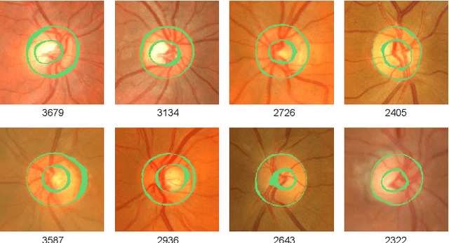
Abstract:Optic disc and cup segmentation play a crucial role in automating the screening and diagnosis of optic glaucoma. While data-driven convolutional neural networks (CNNs) show promise in this area, the inherent ambiguity of segmenting object and background boundaries in the task of optic disc and cup segmentation leads to noisy annotations that impact model performance. To address this, we propose an innovative label-denoising method of Multiple Pseudo-labels Noise-aware Network (MPNN) for accurate optic disc and cup segmentation. Specifically, the Multiple Pseudo-labels Generation and Guided Denoising (MPGGD) module generates pseudo-labels by multiple different initialization networks trained on true labels, and the pixel-level consensus information extracted from these pseudo-labels guides to differentiate clean pixels from noisy pixels. The training framework of the MPNN is constructed by a teacher-student architecture to learn segmentation from clean pixels and noisy pixels. Particularly, such a framework adeptly leverages (i) reliable and fundamental insights from clean pixels and (ii) the supplementary knowledge within noisy pixels via multiple perturbation-based unsupervised consistency. Compared to other label-denoising methods, comprehensive experimental results on the RIGA dataset demonstrate our method's excellent performance and significant denoising ability.
PMP-Swin: Multi-Scale Patch Message Passing Swin Transformer for Retinal Disease Classification
Nov 20, 2023Abstract:Retinal disease is one of the primary causes of visual impairment, and early diagnosis is essential for preventing further deterioration. Nowadays, many works have explored Transformers for diagnosing diseases due to their strong visual representation capabilities. However, retinal diseases exhibit milder forms and often present with overlapping signs, which pose great difficulties for accurate multi-class classification. Therefore, we propose a new framework named Multi-Scale Patch Message Passing Swin Transformer for multi-class retinal disease classification. Specifically, we design a Patch Message Passing (PMP) module based on the Message Passing mechanism to establish global interaction for pathological semantic features and to exploit the subtle differences further between different diseases. Moreover, considering the various scale of pathological features we integrate multiple PMP modules for different patch sizes. For evaluation, we have constructed a new dataset, named OPTOS dataset, consisting of 1,033 high-resolution fundus images photographed by Optos camera and conducted comprehensive experiments to validate the efficacy of our proposed method. And the results on both the public dataset and our dataset demonstrate that our method achieves remarkable performance compared to state-of-the-art methods.
MSE-Nets: Multi-annotated Semi-supervised Ensemble Networks for Improving Segmentation of Medical Image with Ambiguous Boundaries
Nov 17, 2023


Abstract:Medical image segmentation annotations exhibit variations among experts due to the ambiguous boundaries of segmented objects and backgrounds in medical images. Although using multiple annotations for each image in the fully-supervised has been extensively studied for training deep models, obtaining a large amount of multi-annotated data is challenging due to the substantial time and manpower costs required for segmentation annotations, resulting in most images lacking any annotations. To address this, we propose Multi-annotated Semi-supervised Ensemble Networks (MSE-Nets) for learning segmentation from limited multi-annotated and abundant unannotated data. Specifically, we introduce the Network Pairwise Consistency Enhancement (NPCE) module and Multi-Network Pseudo Supervised (MNPS) module to enhance MSE-Nets for the segmentation task by considering two major factors: (1) to optimize the utilization of all accessible multi-annotated data, the NPCE separates (dis)agreement annotations of multi-annotated data at the pixel level and handles agreement and disagreement annotations in different ways, (2) to mitigate the introduction of imprecise pseudo-labels, the MNPS extends the training data by leveraging consistent pseudo-labels from unannotated data. Finally, we improve confidence calibration by averaging the predictions of base networks. Experiments on the ISIC dataset show that we reduced the demand for multi-annotated data by 97.75\% and narrowed the gap with the best fully-supervised baseline to just a Jaccard index of 4\%. Furthermore, compared to other semi-supervised methods that rely only on a single annotation or a combined fusion approach, the comprehensive experimental results on ISIC and RIGA datasets demonstrate the superior performance of our proposed method in medical image segmentation with ambiguous boundaries.
Learning from Noisy Labels Generated by Extremely Point Annotations for OCT Fluid Segmentation
Jun 05, 2023


Abstract:Automatic segmentation of fluid in OCT (Optical Coherence Tomography) images is beneficial for ophthalmologists to make an accurate diagnosis. Currently, data-driven convolutional neural networks (CNNs) have achieved great success in OCT fluid segmentation. However, obtaining pixel-level masks of OCT images is time-consuming and requires expertise. The popular weakly-supervised strategy is to generate noisy pseudo-labels from weak annotations, but the noise information introduced may mislead the model training. To address this issue, (i) we propose a superpixel-guided method for generating noisy labels from weak point annotations, called Point to Noisy by Superpixel (PNS), which limits the network from over-fitting noise by assigning low confidence to suspiciously noisy label pixels, and (ii) we propose a Two-Stage Mean-Teacher-assisted Confident Learning (2SMTCL) method based on MTCL for multi-category OCT fluid segmentation, which alleviates the uncertainty and computing power consumption introduced by the real-time characterization noise of MTCL. For evaluation, we have constructed a 2D OCT fluid segmentation dataset. Compared with other state-of-art label-denoising methods, comprehensive experimental results demonstrate that the proposed method can achieve excellent performance in OCT fluid segmentation as well as label denoising. Our study provides an efficient, accurate, and practical solution for fluid segmentation of OCT images, which is expected to have a positive impact on the diagnosis and treatment of patients in the field of ophthalmology.
 Add to Chrome
 Add to Firefox
 Add to Edge