Yue-Min Zhu
Relationship between pulmonary nodule malignancy and surrounding pleurae, airways and vessels: a quantitative study using the public LIDC-IDRI dataset
Jun 24, 2021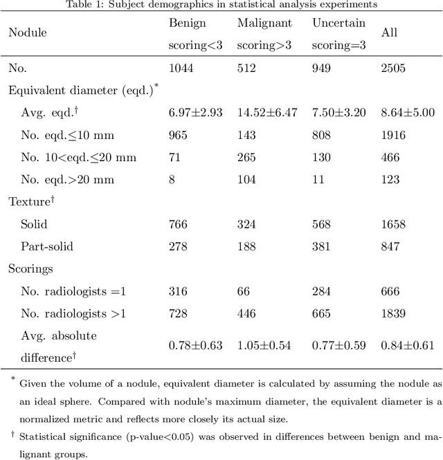
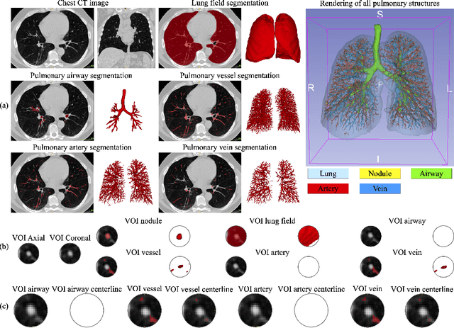
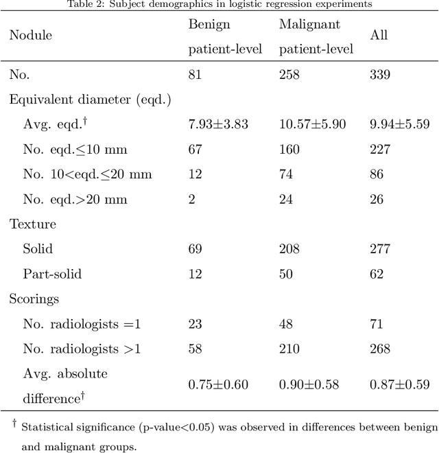
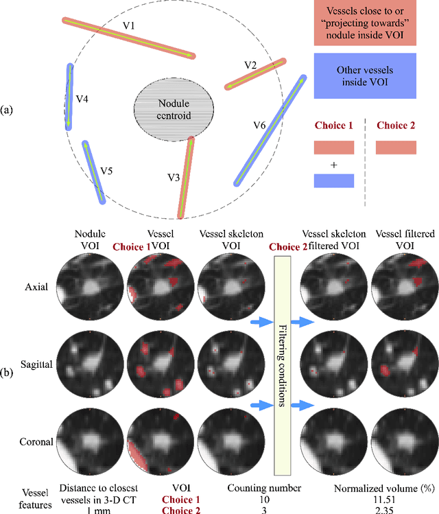
Abstract:To investigate whether the pleurae, airways and vessels surrounding a nodule on non-contrast computed tomography (CT) can discriminate benign and malignant pulmonary nodules. The LIDC-IDRI dataset, one of the largest publicly available CT database, was exploited for study. A total of 1556 nodules from 694 patients were involved in statistical analysis, where nodules with average scorings <3 and >3 were respectively denoted as benign and malignant. Besides, 339 nodules from 113 patients with diagnosis ground-truth were independently evaluated. Computer algorithms were developed to segment pulmonary structures and quantify the distances to pleural surface, airways and vessels, as well as the counting number and normalized volume of airways and vessels near a nodule. Odds ratio (OR) and Chi-square (\chi^2) testing were performed to demonstrate the correlation between features of surrounding structures and nodule malignancy. A non-parametric receiver operating characteristic (ROC) analysis was conducted in logistic regression to evaluate discrimination ability of each structure. For benign and malignant groups, the average distances from nodules to pleural surface, airways and vessels are respectively (6.56, 5.19), (37.08, 26.43) and (1.42, 1.07) mm. The correlation between nodules and the counting number of airways and vessels that contact or project towards nodules are respectively (OR=22.96, \chi^2=105.04) and (OR=7.06, \chi^2=290.11). The correlation between nodules and the volume of airways and vessels are (OR=9.19, \chi^2=159.02) and (OR=2.29, \chi^2=55.89). The areas-under-curves (AUCs) for pleurae, airways and vessels are respectively 0.5202, 0.6943 and 0.6529. Our results show that malignant nodules are often surrounded by more pulmonary structures compared with benign ones, suggesting that features of these structures could be viewed as lung cancer biomarkers.
Learning Tubule-Sensitive CNNs for Pulmonary Airway and Artery-Vein Segmentation in CT
Dec 10, 2020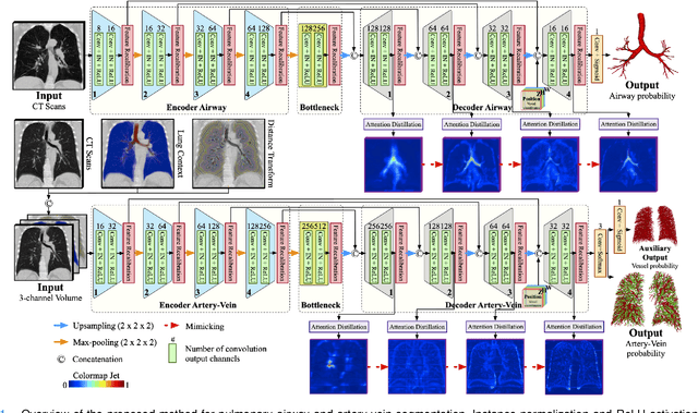
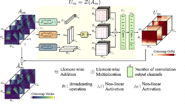
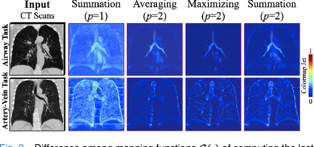
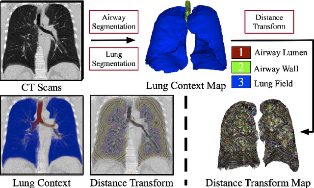
Abstract:Training convolutional neural networks (CNNs) for segmentation of pulmonary airway, artery, and vein is challenging due to sparse supervisory signals caused by the severe class imbalance between tubular targets and background. We present a CNNs-based method for accurate airway and artery-vein segmentation in non-contrast computed tomography. It enjoys superior sensitivity to tenuous peripheral bronchioles, arterioles, and venules. The method first uses a feature recalibration module to make the best use of features learned from the neural networks. Spatial information of features is properly integrated to retain relative priority of activated regions, which benefits the subsequent channel-wise recalibration. Then, attention distillation module is introduced to reinforce representation learning of tubular objects. Fine-grained details in high-resolution attention maps are passing down from one layer to its previous layer recursively to enrich context. Anatomy prior of lung context map and distance transform map is designed and incorporated for better artery-vein differentiation capacity. Extensive experiments demonstrated considerable performance gains brought by these components. Compared with state-of-the-art methods, our method extracted much more branches while maintaining competitive overall segmentation performance. Codes and models will be available later at http://www.pami.sjtu.edu.cn.
AirwayNet: A Voxel-Connectivity Aware Approach for Accurate Airway Segmentation Using Convolutional Neural Networks
Jul 16, 2019
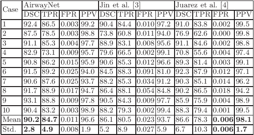


Abstract:Airway segmentation on CT scans is critical for pulmonary disease diagnosis and endobronchial navigation. Manual extraction of airway requires strenuous efforts due to the complicated structure and various appearance of airway. For automatic airway extraction, convolutional neural networks (CNNs) based methods have recently become the state-of-the-art approach. However, there still remains a challenge for CNNs to perceive the tree-like pattern and comprehend the connectivity of airway. To address this, we propose a voxel-connectivity aware approach named AirwayNet for accurate airway segmentation. By connectivity modeling, conventional binary segmentation task is transformed into 26 tasks of connectivity prediction. Thus, our AirwayNet learns both airway structure and relationship between neighboring voxels. To take advantage of context knowledge, lung distance map and voxel coordinates are fed into AirwayNet as additional semantic information. Compared to existing approaches, AirwayNet achieved superior performance, demonstrating the effectiveness of the network's awareness of voxel connectivity.
Varifocal-Net: A Chromosome Classification Approach using Deep Convolutional Networks
Oct 20, 2018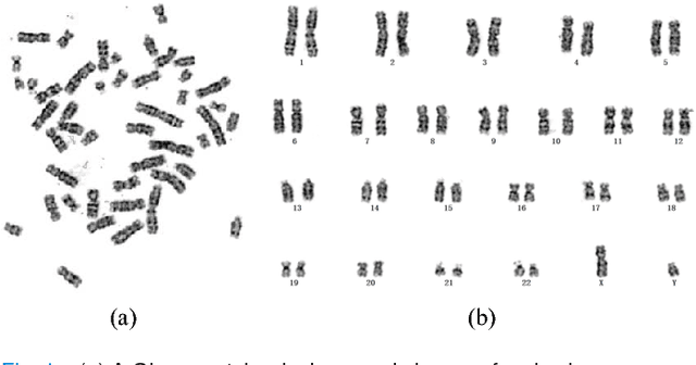
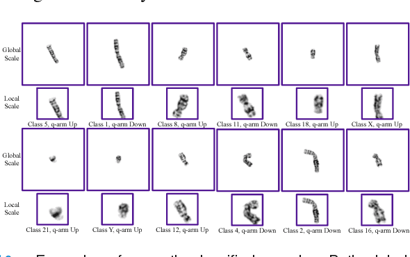
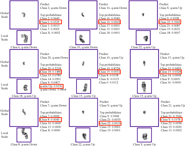
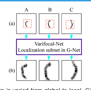
Abstract:Chromosome classification is critical for karyotyping in abnormality diagnosis. To expedite diagnosis process, we present a novel method named Varifocal-Net for simultaneous classification of chromosome's type and polarity using deep convolutional networks. The approach consists of one global-scale network (G-Net) and one local-scale network (L-Net). It follows two stages. The first stage is to learn both global and local features. We extract global features and detect finer local regions via the G-Net. With the proposed varifocal mechanism, we zoom into local parts and extract local features via the L-Net. Residual learning and multi-task learning strategies are utilized to promote high-level feature extraction. The detection of discriminative local parts is fulfilled by a localization subnet of the G-Net, whose training process involves both supervised and weekly-supervised learning. The second stage is to build two multi-layer perceptron classifiers that exploit features of both two scales to boost classification performance. Evaluation results from 1909 karyotyping cases demonstrate that our Varifocal-Net achieved the highest accuracy of 0.9805, 0.9909 and average F1-score of 0.9771, 0.9909 for the type and polarity task, respectively. It outperformed state-of-the-art methods, demonstrating the effectiveness of our Varifocal mechanism and multi-scale feature ensemble.
 Add to Chrome
 Add to Firefox
 Add to Edge