Yangxin Xu
When Gradient Optimization Is Not Enough: $\dagger$ Dispersive and Anchoring Geometric Regularizer for Multimodal Learning
Jan 29, 2026Abstract:Multimodal learning aims to integrate complementary information from heterogeneous modalities, yet strong optimization alone does not guaranty well-structured representations. Even under carefully balanced training schemes, multimodal models often exhibit geometric pathologies, including intra-modal representation collapse and sample-level cross-modal inconsistency, which degrade both unimodal robustness and multimodal fusion. We identify representation geometry as a missing control axis in multimodal learning and propose \regName, a lightweight geometry-aware regularization framework. \regName enforces two complementary constraints on intermediate embeddings: an intra-modal dispersive regularization that promotes representation diversity, and an inter-modal anchoring regularization that bounds sample-level cross-modal drift without rigid alignment. The proposed regularizer is plug-and-play, requires no architectural modifications, and is compatible with various training paradigms. Extensive experiments across multiple multimodal benchmarks demonstrate consistent improvements in both multimodal and unimodal performance, showing that explicitly regulating representation geometry effectively mitigates modality trade-offs.
Style Transfer Enabled Sim2Real Framework for Efficient Learning of Robotic Ultrasound Image Analysis Using Simulated Data
May 16, 2023Abstract:Robotic ultrasound (US) systems have shown great potential to make US examinations easier and more accurate. Recently, various machine learning techniques have been proposed to realize automatic US image interpretation for robotic US acquisition tasks. However, obtaining large amounts of real US imaging data for training is usually expensive or even unfeasible in some clinical applications. An alternative is to build a simulator to generate synthetic US data for training, but the differences between simulated and real US images may result in poor model performance. This work presents a Sim2Real framework to efficiently learn robotic US image analysis tasks based only on simulated data for real-world deployment. A style transfer module is proposed based on unsupervised contrastive learning and used as a preprocessing step to convert the real US images into the simulation style. Thereafter, a task-relevant model is designed to combine CNNs with vision transformers to generate the task-dependent prediction with improved generalization ability. We demonstrate the effectiveness of our method in an image regression task to predict the probe position based on US images in robotic transesophageal echocardiography (TEE). Our results show that using only simulated US data and a small amount of unlabelled real data for training, our method can achieve comparable performance to semi-supervised and fully supervised learning methods. Moreover, the effectiveness of our previously proposed CT-based US image simulation method is also indirectly confirmed.
Closed-Loop Magnetic Manipulation for Robotic Transesophageal Echocardiography
Jan 16, 2023


Abstract:This paper presents a closed-loop magnetic manipulation framework for robotic transesophageal echocardiography (TEE) acquisitions. Different from previous work on intracorporeal robotic ultrasound acquisitions that focus on continuum robot control, we first investigate the use of magnetic control methods for more direct, intuitive, and accurate manipulation of the distal tip of the probe. We modify a standard TEE probe by attaching a permanent magnet and an inertial measurement unit sensor to the probe tip and replacing the flexible gastroscope with a soft tether containing only wires for transmitting ultrasound signals, and show that 6-DOF localization and 5-DOF closed-loop control of the probe can be achieved with an external permanent magnet based on the fusion of internal inertial measurement and external magnetic field sensing data. The proposed method does not require complex structures or motions of the actuator and the probe compared with existing magnetic manipulation methods. We have conducted extensive experiments to validate the effectiveness of the framework in terms of localization accuracy, update rate, workspace size, and tracking accuracy. In addition, our results obtained on a realistic cardiac tissue-mimicking phantom show that the proposed framework is applicable in real conditions and can generally meet the requirements for tele-operated TEE acquisitions.
Image-Guided Navigation of a Robotic Ultrasound Probe for Autonomous Spinal Sonography Using a Shadow-aware Dual-Agent Framework
Nov 10, 2021


Abstract:Ultrasound (US) imaging is commonly used to assist in the diagnosis and interventions of spine diseases, while the standardized US acquisitions performed by manually operating the probe require substantial experience and training of sonographers. In this work, we propose a novel dual-agent framework that integrates a reinforcement learning (RL) agent and a deep learning (DL) agent to jointly determine the movement of the US probe based on the real-time US images, in order to mimic the decision-making process of an expert sonographer to achieve autonomous standard view acquisitions in spinal sonography. Moreover, inspired by the nature of US propagation and the characteristics of the spinal anatomy, we introduce a view-specific acoustic shadow reward to utilize the shadow information to implicitly guide the navigation of the probe toward different standard views of the spine. Our method is validated in both quantitative and qualitative experiments in a simulation environment built with US data acquired from 17 volunteers. The average navigation accuracy toward different standard views achieves 5.18mm/5.25deg and 12.87mm/17.49deg in the intra- and inter-subject settings, respectively. The results demonstrate that our method can effectively interpret the US images and navigate the probe to acquire multiple standard views of the spine.
Autonomous Magnetic Navigation Framework for Active Wireless Capsule Endoscopy Inspired by Conventional Colonoscopy Procedures
Nov 03, 2021


Abstract:In recent years, simultaneous magnetic actuation and localization (SMAL) for active wireless capsule endoscopy (WCE) has been intensively studied to improve the efficiency and accuracy of the examination. In this paper, we propose an autonomous magnetic navigation framework for active WCE that mimics the "insertion" and "withdrawal" procedures performed by an expert physician in conventional colonoscopy, thereby enabling efficient and accurate navigation of a robotic capsule endoscope in the intestine with minimal user effort. First, the capsule is automatically propelled through the unknown intestinal environment and generate a viable path to represent the environment. Then, the capsule is autonomously navigated towards any point selected on the intestinal trajectory to allow accurate and repeated inspections of suspicious lesions. Moreover, we implement the navigation framework on a robotic system incorporated with advanced SMAL algorithms, and validate it in the navigation in various tubular environments using phantoms and an ex-vivo pig colon. Our results demonstrate that the proposed autonomous navigation framework can effectively navigate the capsule in unknown, complex tubular environments with a satisfactory accuracy, repeatability and efficiency compared with manual operation.
Automatic Recognition of Abdominal Organs in Ultrasound Images based on Deep Neural Networks and K-Nearest-Neighbor Classification
Oct 09, 2021


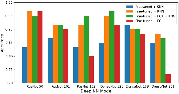
Abstract:Abdominal ultrasound imaging has been widely used to assist in the diagnosis and treatment of various abdominal organs. In order to shorten the examination time and reduce the cognitive burden on the sonographers, we present a classification method that combines the deep learning techniques and k-Nearest-Neighbor (k-NN) classification to automatically recognize various abdominal organs in the ultrasound images in real time. Fine-tuned deep neural networks are used in combination with PCA dimension reduction to extract high-level features from raw ultrasound images, and a k-NN classifier is employed to predict the abdominal organ in the image. We demonstrate the effectiveness of our method in the task of ultrasound image classification to automatically recognize six abdominal organs. A comprehensive comparison of different configurations is conducted to study the influence of different feature extractors and classifiers on the classification accuracy. Both quantitative and qualitative results show that with minimal training effort, our method can "lazily" recognize the abdominal organs in the ultrasound images in real time with an accuracy of 96.67%. Our implementation code is publicly available at: https://github.com/LeeKeyu/abdominal_ultrasound_classification.
Trajectory Following of a Reciprocally Rotating Magnetic Capsule in a Tubular Environment
Aug 26, 2021


Abstract:Currently used wireless capsule endoscopy (WCE) is limited in terms of inspection time and flexibility since the capsule is passively moved by peristalsis and cannot be accurately positioned. Different methods have been proposed to facilitate active locomotion of WCE based on simultaneous magnetic actuation and localization technologies. In this work, we first investigate the trajectory following (TF) problem of a robotic capsule under reciprocally rotating magnetic actuation in a tubular environment, in order to safely, efficiently and accurately perform inspection of the intestine at given points. Specifically, four TF strategies are proposed and analyzed based on the PD controller, adaptive controller, model predictive controller and robust multi-stage model predictive controller. Moreover, we take into consideration the intestinal peristalsis and friction in our controller design by explicitly modeling the interdigestive migrating motor complex (MMC) mechanism in the intestine. We validate our methods in real-world experiments in various tubular environments including phantoms with different shapes and an ex-vivo pig colon. The results show that our approach can effectively actuate a reciprocally rotating capsule to follow a desired trajectory in complex tubular environments, thereby enabling repeated and accurate inspection of the intestine for high-quality diagnosis.
Adaptive Simultaneous Magnetic Actuation and Localization for WCE in a Tubular Environment
Aug 25, 2021


Abstract:Simultaneous Magnetic Actuation and Localization (SMAL) is a promising technology for active wireless capsule endoscopy (WCE). In this paper, an adaptive SMAL system is presented to efficiently propel and precisely locate a capsule in a tubular environment with complex shapes. In order to track the capsule with high localization accuracy and update frequency in a large workspace, we propose a mechanism that can automatically activate a sub-array of sensors with the optimal layout during the capsule movement. The improved multiple objects tracking (IMOT) method is simplified and adapted to our system to estimate the 6-D pose of the capsule in real time. Also, we study the locomotion of a magnetically actuated capsule in a tubular environment, and formulate a method to adaptively adjust the pose of the actuator to improve the propulsion efficiency. Our presented methods are applicable to other permanent magnet-based SMAL systems, and help to improve the actuation efficiency of active WCE. We verify the effectiveness of our proposed system in extensive experiments on phantoms and ex-vivo animal organs. The results demonstrate that our system can achieve convincing performance compared with the state-of-the-art ones in terms of actuation efficiency, workspace size, robustness, localization accuracy and update frequency.
On Reciprocally Rotating Magnetic Actuation of a Robotic Capsule in Unknown Tubular Environments
Aug 25, 2021


Abstract:Active wireless capsule endoscopy (WCE) based on simultaneous magnetic actuation and localization (SMAL) techniques holds great promise for improving diagnostic accuracy, reducing examination time and relieving operator burden. To date, the rotating magnetic actuation methods have been constrained to use a continuously rotating permanent magnet. In this paper, we first propose the reciprocally rotating magnetic actuation (RRMA) approach for active WCE to enhance patient safety. We first show how to generate a desired reciprocally rotating magnetic field for capsule actuation, and provide a theoretical analysis of the potential risk of causing volvulus due to the capsule motion. Then, an RRMA-based SMAL workflow is presented to automatically propel a capsule in an unknown tubular environment. We validate the effectiveness of our method in real-world experiments to automatically propel a robotic capsule in an ex-vivo pig colon. The experiment results show that our approach can achieve efficient and robust propulsion of the capsule with an average moving speed of $2.48 mm/s$ in the pig colon, and demonstrate the potential of using RRMA to enhance patient safety, reduce the inspection time, and improve the clinical acceptance of this technology.
Autonomous Navigation of an Ultrasound Probe Towards Standard Scan Planes with Deep Reinforcement Learning
Mar 01, 2021


Abstract:Autonomous ultrasound (US) acquisition is an important yet challenging task, as it involves interpretation of the highly complex and variable images and their spatial relationships. In this work, we propose a deep reinforcement learning framework to autonomously control the 6-D pose of a virtual US probe based on real-time image feedback to navigate towards the standard scan planes under the restrictions in real-world US scans. Furthermore, we propose a confidence-based approach to encode the optimization of image quality in the learning process. We validate our method in a simulation environment built with real-world data collected in the US imaging of the spine. Experimental results demonstrate that our method can perform reproducible US probe navigation towards the standard scan plane with an accuracy of $4.91mm/4.65^\circ$ in the intra-patient setting, and accomplish the task in the intra- and inter-patient settings with a success rate of $92\%$ and $46\%$, respectively. The results also show that the introduction of image quality optimization in our method can effectively improve the navigation performance.
 Add to Chrome
 Add to Firefox
 Add to Edge