William V. Stoecker
Increasing Melanoma Diagnostic Confidence: Forcing the Convolutional Network to Learn from the Lesion
May 16, 2023Abstract:Deep learning implemented with convolutional network architectures can exceed specialists' diagnostic accuracy. However, whole-image deep learning trained on a given dataset may not generalize to other datasets. The problem arises because extra-lesional features - ruler marks, ink marks, and other melanoma correlates - may serve as information leaks. These extra-lesional features, discoverable by heat maps, degrade melanoma diagnostic performance and cause techniques learned on one data set to fail to generalize. We propose a novel technique to improve melanoma recognition by an EfficientNet model. The model trains the network to detect the lesion and learn features from the detected lesion. A generalizable elliptical segmentation model for lesions was developed, with an ellipse enclosing a lesion and the ellipse enclosed by an extended rectangle (bounding box). The minimal bounding box was extended by 20% to allow some background around the lesion. The publicly available International Skin Imaging Collaboration (ISIC) 2020 skin lesion image dataset was used to evaluate the effectiveness of the proposed method. Our test results show that the proposed method improved diagnostic accuracy by increasing the mean area under receiver operating characteristic curve (mean AUC) score from 0.9 to 0.922. Additionally, correctly diagnosed scores are also improved, providing better separation of scores, thereby increasing melanoma diagnostic confidence. The proposed lesion-focused convolutional technique warrants further study.
Feature based Sequential Classifier with Attention Mechanism
Jul 22, 2020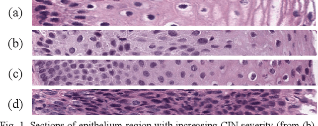
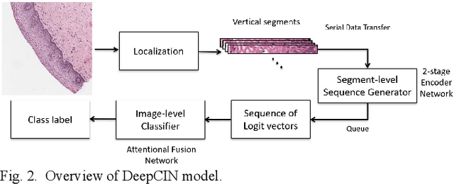
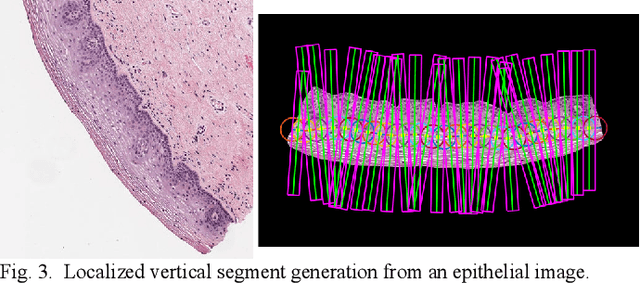
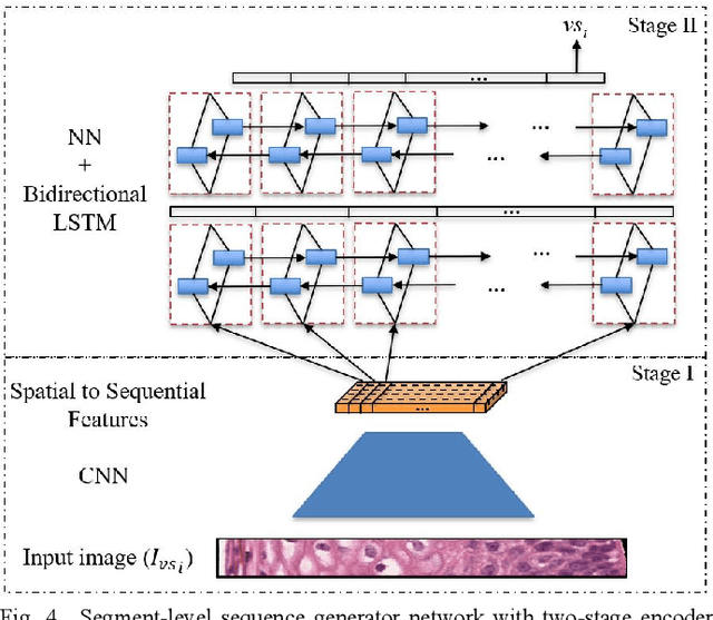
Abstract:Cervical cancer is one of the deadliest cancers affecting women globally. Cervical intraepithelial neoplasia (CIN) assessment using histopathological examination of cervical biopsy slides is subject to interobserver variability. Automated processing of digitized histopathology slides has the potential for more accurate classification for CIN grades from normal to increasing grades of pre-malignancy: CIN1, CIN2 and CIN3. Cervix disease is generally understood to progress from the bottom (basement membrane) to the top of the epithelium. To model this relationship of disease severity to spatial distribution of abnormalities, we propose a network pipeline, DeepCIN, to analyze high-resolution epithelium images (manually extracted from whole-slide images) hierarchically by focusing on localized vertical regions and fusing this local information for determining Normal/CIN classification. The pipeline contains two classifier networks: 1) a cross-sectional, vertical segment-level sequence generator (two-stage encoder model) is trained using weak supervision to generate feature sequences from the vertical segments to preserve the bottom-to-top feature relationships in the epithelium image data; 2) an attention-based fusion network image-level classifier predicting the final CIN grade by merging vertical segment sequences. The model produces the CIN classification results and also determines the vertical segment contributions to CIN grade prediction. Experiments show that DeepCIN achieves pathologist-level CIN classification accuracy.
Lesion Border Detection in Dermoscopy Images
Oct 30, 2010


Abstract:Background: Dermoscopy is one of the major imaging modalities used in the diagnosis of melanoma and other pigmented skin lesions. Due to the difficulty and subjectivity of human interpretation, computerized analysis of dermoscopy images has become an important research area. One of the most important steps in dermoscopy image analysis is the automated detection of lesion borders. Methods: In this article, we present a systematic overview of the recent border detection methods in the literature paying particular attention to computational issues and evaluation aspects. Conclusion: Common problems with the existing approaches include the acquisition, size, and diagnostic distribution of the test image set, the evaluation of the results, and the inadequate description of the employed methods. Border determination by dermatologists appears to depend upon higher-level knowledge, therefore it is likely that the incorporation of domain knowledge in automated methods will enable them to perform better, especially in sets of images with a variety of diagnoses.
* 10 pages, 1 figure, 3 tables
Approximate Lesion Localization in Dermoscopy Images
Sep 06, 2010


Abstract:Background: Dermoscopy is one of the major imaging modalities used in the diagnosis of melanoma and other pigmented skin lesions. Due to the difficulty and subjectivity of human interpretation, automated analysis of dermoscopy images has become an important research area. Border detection is often the first step in this analysis. Methods: In this article, we present an approximate lesion localization method that serves as a preprocessing step for detecting borders in dermoscopy images. In this method, first the black frame around the image is removed using an iterative algorithm. The approximate location of the lesion is then determined using an ensemble of thresholding algorithms. Results: The method is tested on a set of 428 dermoscopy images. The localization error is quantified by a metric that uses dermatologist determined borders as the ground truth. Conclusion: The results demonstrate that the method presented here achieves both fast and accurate localization of lesions in dermoscopy images.
An Improved Objective Evaluation Measure for Border Detection in Dermoscopy Images
Sep 06, 2010


Abstract:Background: Dermoscopy is one of the major imaging modalities used in the diagnosis of melanoma and other pigmented skin lesions. Due to the difficulty and subjectivity of human interpretation, dermoscopy image analysis has become an important research area. One of the most important steps in dermoscopy image analysis is the automated detection of lesion borders. Although numerous methods have been developed for the detection of lesion borders, very few studies were comprehensive in the evaluation of their results. Methods: In this paper, we evaluate five recent border detection methods on a set of 90 dermoscopy images using three sets of dermatologist-drawn borders as the ground-truth. In contrast to previous work, we utilize an objective measure, the Normalized Probabilistic Rand Index, which takes into account the variations in the ground-truth images. Conclusion: The results demonstrate that the differences between four of the evaluated border detection methods are in fact smaller than those predicted by the commonly used XOR measure.
Automatic Detection of Blue-White Veil and Related Structures in Dermoscopy Images
Sep 06, 2010
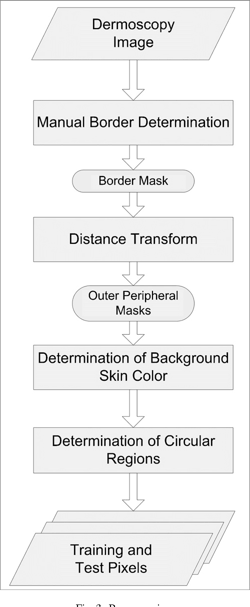
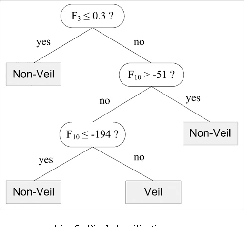
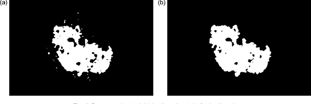
Abstract:Dermoscopy is a non-invasive skin imaging technique, which permits visualization of features of pigmented melanocytic neoplasms that are not discernable by examination with the naked eye. One of the most important features for the diagnosis of melanoma in dermoscopy images is the blue-white veil (irregular, structureless areas of confluent blue pigmentation with an overlying white "ground-glass" film). In this article, we present a machine learning approach to the detection of blue-white veil and related structures in dermoscopy images. The method involves contextual pixel classification using a decision tree classifier. The percentage of blue-white areas detected in a lesion combined with a simple shape descriptor yielded a sensitivity of 69.35% and a specificity of 89.97% on a set of 545 dermoscopy images. The sensitivity rises to 78.20% for detection of blue veil in those cases where it is a primary feature for melanoma recognition.
 Add to Chrome
 Add to Firefox
 Add to Edge