Stefano Moriconi
Deep generative computed perfusion-deficit mapping of ischaemic stroke
Feb 03, 2025


Abstract:Focal deficits in ischaemic stroke result from impaired perfusion downstream of a critical vascular occlusion. While parenchymal lesions are traditionally used to predict clinical deficits, the underlying pattern of disrupted perfusion provides information upstream of the lesion, potentially yielding earlier predictive and localizing signals. Such perfusion maps can be derived from routine CT angiography (CTA) widely deployed in clinical practice. Analysing computed perfusion maps from 1,393 CTA-imaged-patients with acute ischaemic stroke, we use deep generative inference to localise neural substrates of NIHSS sub-scores. We show that our approach replicates known lesion-deficit relations without knowledge of the lesion itself and reveals novel neural dependents. The high achieved anatomical fidelity suggests acute CTA-derived computed perfusion maps may be of substantial clinical-and-scientific value in rich phenotyping of acute stroke. Using only hyperacute imaging, deep generative inference could power highly expressive models of functional anatomical relations in ischaemic stroke within the pre-interventional window.
Framework to generate perfusion map from CT and CTA images in patients with acute ischemic stroke: A longitudinal and cross-sectional study
Apr 05, 2024


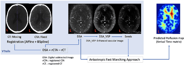
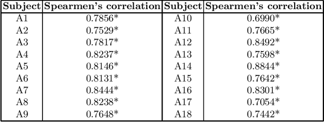
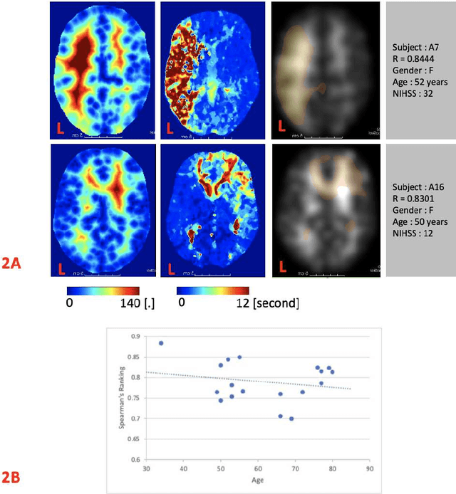
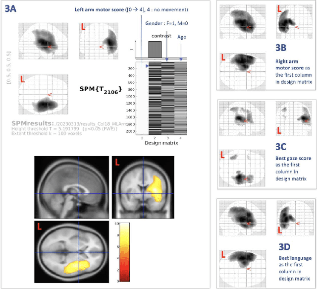
Abstract:Stroke is a leading cause of disability and death. Effective treatment decisions require early and informative vascular imaging. 4D perfusion imaging is ideal but rarely available within the first hour after stroke, whereas plain CT and CTA usually are. Hence, we propose a framework to extract a predicted perfusion map (PPM) derived from CT and CTA images. In all eighteen patients, we found significantly high spatial similarity (with average Spearman's correlation = 0.7893) between our predicted perfusion map (PPM) and the T-max map derived from 4D-CTP. Voxelwise correlations between the PPM and National Institutes of Health Stroke Scale (NIHSS) subscores for L/R hand motor, gaze, and language on a large cohort of 2,110 subjects reliably mapped symptoms to expected infarct locations. Therefore our PPM could serve as an alternative for 4D perfusion imaging, if the latter is unavailable, to investigate blood perfusion in the first hours after hospital admission.
Near-Real-Time Mueller Polarimetric Image Processing for Neurosurgical Intervention
Mar 01, 2024Abstract:Wide-field imaging Mueller polarimetry is a revolutionary, label-free, and non-invasive modality for computer-aided intervention: in neurosurgery it aims to provide visual feedback of white matter fibre bundle orientation from derived parameters. Conventionally, robust polarimetric parameters are estimated after averaging multiple measurements of intensity for each pair of probing and detected polarised light. Long multi-shot averaging, however, is not compatible with real-time in-vivo imaging, and the current performance of polarimetric data processing hinders the translation to clinical practice. A learning-based denoising framework is tailored for fast, single-shot, noisy acquisitions of polarimetric intensities. Also, performance-optimised image processing tools are devised for the derivation of clinically relevant parameters. The combination recovers accurate polarimetric parameters from fast acquisitions with near-real-time performance, under the assumption of pseudo-Gaussian polarimetric acquisition noise. The denoising framework is trained, validated, and tested on experimental data comprising tumour-free and diseased human brain samples in different conditions. Accuracy and image quality indices showed significant improvements on testing data for a fast single-pass denoising versus the state-of-the-art and high polarimetric image quality standards. The computational time is reported for the end-to-end processing. The end-to-end image processing achieved real-time performance for a localised field of view. The denoised polarimetric intensities produced visibly clear directional patterns of neuronal fibre tracts in line with reference polarimetric image quality standards; directional disruption was kept in case of neoplastic lesions. The presented advances pave the way towards feasible oncological neurosurgical translations of novel, label free, interventional feedback.
Elastic Registration of Geodesic Vascular Graphs
Sep 14, 2018


Abstract:Vascular graphs can embed a number of high-level features, from morphological parameters, to functional biomarkers, and represent an invaluable tool for longitudinal and cross-sectional clinical inference. This, however, is only feasible when graphs are co-registered together, allowing coherent multiple comparisons. The robust registration of vascular topologies stands therefore as key enabling technology for group-wise analyses. In this work, we present an end-to-end vascular graph registration approach, that aligns networks with non-linear geometries and topological deformations, by introducing a novel overconnected geodesic vascular graph formulation, and without enforcing any anatomical prior constraint. The 3D elastic graph registration is then performed with state-of-the-art graph matching methods used in computer vision. Promising results of vascular matching are found using graphs from synthetic and real angiographies. Observations and future designs are discussed towards potential clinical applications.
VTrails: Inferring Vessels with Geodesic Connectivity Trees
Jun 08, 2018


Abstract:The analysis of vessel morphology and connectivity has an impact on a number of cardiovascular and neurovascular applications by providing patient-specific high-level quantitative features such as spatial location, direction and scale. In this paper we present an end-to-end approach to extract an acyclic vascular tree from angiographic data by solving a connectivity-enforcing anisotropic fast marching over a voxel-wise tensor field representing the orientation of the underlying vascular tree. The method is validated using synthetic and real vascular images. We compare VTrails against classical and state-of-the-art ridge detectors for tubular structures by assessing the connectedness of the vesselness map and inspecting the synthesized tensor field as proof of concept. VTrails performance is evaluated on images with different levels of degradation: we verify that the extracted vascular network is an acyclic graph (i.e. a tree), and we report the extraction accuracy, precision and recall.
 Add to Chrome
 Add to Firefox
 Add to Edge