Stefan Maetschke
Understanding in Artificial Intelligence
Jan 17, 2021
Abstract:Current Artificial Intelligence (AI) methods, most based on deep learning, have facilitated progress in several fields, including computer vision and natural language understanding. The progress of these AI methods is measured using benchmarks designed to solve challenging tasks, such as visual question answering. A question remains of how much understanding is leveraged by these methods and how appropriate are the current benchmarks to measure understanding capabilities. To answer these questions, we have analysed existing benchmarks and their understanding capabilities, defined by a set of understanding capabilities, and current research streams. We show how progress has been made in benchmark development to measure understanding capabilities of AI methods and we review as well how current methods develop understanding capabilities.
Inference of visual field test performance from OCT volumes using deep learning
Aug 26, 2019


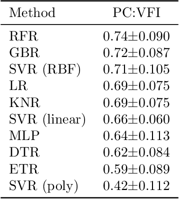
Abstract:Visual field tests (VFT) are pivotal for glaucoma diagnosis and conducted regularly to monitor disease progression. Here we address the question to what degree aggregate VFT measurements such as Visual Field Index (VFI) and Mean Deviation (MD) can be inferred from Optical Coherence Tomography (OCT) scans of the Optic Nerve Head (ONH) or the macula. Accurate inference of VFT measurements from OCT could reduce examination time and cost. We propose a novel 3D Convolutional Neural Network (CNN) for this task and compare its accuracy with classical machine learning (ML) algorithms trained on common, segmentation-based OCT, features employed for glaucoma diagnostics. Peak accuracies were achieved on ONH scans when inferring VFI with a Pearson Correlation (PC) of 0.88$\pm$0.035 for the CNN and a significantly lower (p $<$ 0.01) PC of 0.74$\pm$0.090 for the best performing, classical ML algorithm - a Random Forest regressor. Estimation of MD was equally accurate with a PC of 0.88$\pm$0.023 on ONH scans for the CNN.
A feature agnostic approach for glaucoma detection in OCT volumes
Aug 15, 2018


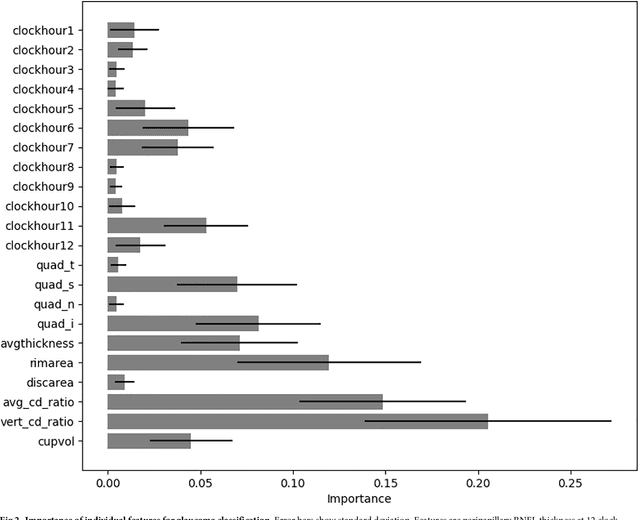
Abstract:Optical coherence tomography (OCT) based measurements of retinal layer thickness, such as the retinal nerve fibre layer (RNFL) and the ganglion cell with inner plexiform layer (GCIPL) are commonly used for the diagnosis and monitoring of glaucoma. Previously, machine learning techniques have utilized segmentation-based imaging features such as the peripapillary RNFL thickness and the cup-to-disc ratio. Here, we propose a deep learning technique that classifies eyes as healthy or glaucomatous directly from raw, unsegmented OCT volumes of the optic nerve head (ONH) using a 3D Convolutional Neural Network (CNN). We compared the accuracy of this technique with various feature-based machine learning algorithms and demonstrated the superiority of the proposed deep learning based method. Logistic regression was found to be the best performing classical machine learning technique with an AUC of 0.89. In direct comparison, the deep learning approach achieved a substantially higher AUC of 0.94 with the additional advantage of providing insight into which regions of an OCT volume are important for glaucoma detection. Computing Class Activation Maps (CAM), we found that the CNN identified neuroretinal rim and optic disc cupping as well as the lamina cribrosa (LC) and its surrounding areas as the regions significantly associated with the glaucoma classification. These regions anatomically correspond to the well established and commonly used clinical markers for glaucoma diagnosis such as increased cup volume, cup diameter, and neuroretinal rim thinning at the superior and inferior segments.
 Add to Chrome
 Add to Firefox
 Add to Edge