 Add to Chrome
 Add to Firefox
 Add to Edge
SeLIP: Similarity Enhanced Contrastive Language Image Pretraining for Multi-modal Head MRI
Mar 25, 2025


Despite that deep learning (DL) methods have presented tremendous potential in many medical image analysis tasks, the practical applications of medical DL models are limited due to the lack of enough data samples with manual annotations. By noting that the clinical radiology examinations are associated with radiology reports that describe the images, we propose to develop a foundation model for multi-model head MRI by using contrastive learning on the images and the corresponding radiology findings. In particular, a contrastive learning framework is proposed, where a mixed syntax and semantic similarity matching metric is integrated to reduce the thirst of extreme large dataset in conventional contrastive learning framework. Our proposed similarity enhanced contrastive language image pretraining (SeLIP) is able to effectively extract more useful features. Experiments revealed that our proposed SeLIP performs well in many downstream tasks including image-text retrieval task, classification task, and image segmentation, which highlights the importance of considering the similarities among texts describing different images in developing medical image foundation models.
Automated Segmentation of Brain Gray Matter Nuclei on Quantitative Susceptibility Mapping Using Deep Convolutional Neural Network
Aug 03, 2020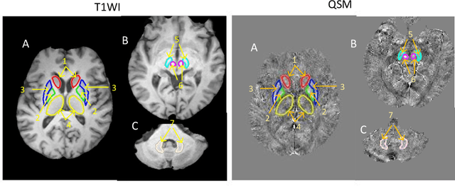
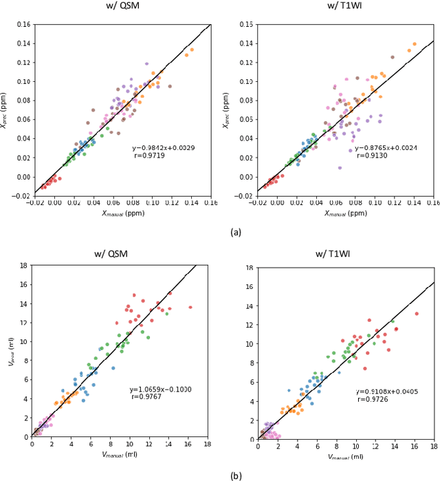
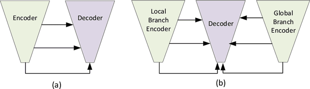
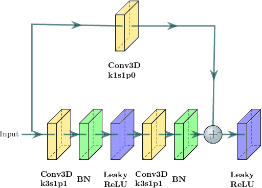
Abnormal iron accumulation in the brain subcortical nuclei has been reported to be correlated to various neurodegenerative diseases, which can be measured through the magnetic susceptibility from the quantitative susceptibility mapping (QSM). To quantitively measure the magnetic susceptibility, the nuclei should be accurately segmented, which is a tedious task for clinicians. In this paper, we proposed a double-branch residual-structured U-Net (DB-ResUNet) based on 3D convolutional neural network (CNN) to automatically segment such brain gray matter nuclei. To better tradeoff between segmentation accuracy and the memory efficiency, the proposed DB-ResUNet fed image patches with high resolution and the patches with low resolution but larger field of view into the local and global branches, respectively. Experimental results revealed that by jointly using QSM and T$_\text{1}$ weighted imaging (T$_\text{1}$WI) as inputs, the proposed method was able to achieve better segmentation accuracy over its single-branch counterpart, as well as the conventional atlas-based method and the classical 3D-UNet structure. The susceptibility values and the volumes were also measured, which indicated that the measurements from the proposed DB-ResUNet are able to present high correlation with values from the manually annotated regions of interest.