Sheheryar Khan
A Layered Self-Supervised Knowledge Distillation Framework for Efficient Multimodal Learning on the Edge
Jun 08, 2025Abstract:We introduce Layered Self-Supervised Knowledge Distillation (LSSKD) framework for training compact deep learning models. Unlike traditional methods that rely on pre-trained teacher networks, our approach appends auxiliary classifiers to intermediate feature maps, generating diverse self-supervised knowledge and enabling one-to-one transfer across different network stages. Our method achieves an average improvement of 4.54\% over the state-of-the-art PS-KD method and a 1.14% gain over SSKD on CIFAR-100, with a 0.32% improvement on ImageNet compared to HASSKD. Experiments on Tiny ImageNet and CIFAR-100 under few-shot learning scenarios also achieve state-of-the-art results. These findings demonstrate the effectiveness of our approach in enhancing model generalization and performance without the need for large over-parameterized teacher networks. Importantly, at the inference stage, all auxiliary classifiers can be removed, yielding no extra computational cost. This makes our model suitable for deploying small language models on affordable low-computing devices. Owing to its lightweight design and adaptability, our framework is particularly suitable for multimodal sensing and cyber-physical environments that require efficient and responsive inference. LSSKD facilitates the development of intelligent agents capable of learning from limited sensory data under weak supervision.
CartiMorph: a framework for automated knee articular cartilage morphometrics
Aug 03, 2023Abstract:We introduce CartiMorph, a framework for automated knee articular cartilage morphometrics. It takes an image as input and generates quantitative metrics for cartilage subregions, including the percentage of full-thickness cartilage loss (FCL), mean thickness, surface area, and volume. CartiMorph leverages the power of deep learning models for hierarchical image feature representation. Deep learning models were trained and validated for tissue segmentation, template construction, and template-to-image registration. We established methods for surface-normal-based cartilage thickness mapping, FCL estimation, and rule-based cartilage parcellation. Our cartilage thickness map showed less error in thin and peripheral regions. We evaluated the effectiveness of the adopted segmentation model by comparing the quantitative metrics obtained from model segmentation and those from manual segmentation. The root-mean-squared deviation of the FCL measurements was less than 8%, and strong correlations were observed for the mean thickness (Pearson's correlation coefficient $\rho \in [0.82,0.97]$), surface area ($\rho \in [0.82,0.98]$) and volume ($\rho \in [0.89,0.98]$) measurements. We compared our FCL measurements with those from a previous study and found that our measurements deviated less from the ground truths. We observed superior performance of the proposed rule-based cartilage parcellation method compared with the atlas-based approach. CartiMorph has the potential to promote imaging biomarkers discovery for knee osteoarthritis.
Multipath CNN with alpha matte inference for knee tissue segmentation from MRI
Sep 29, 2021


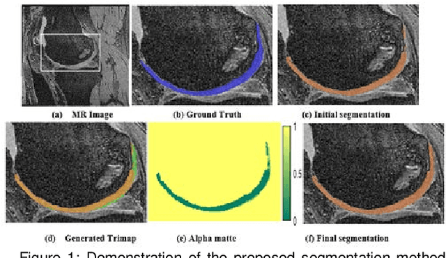
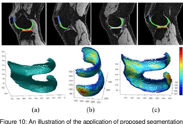
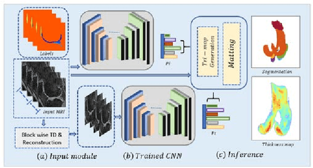
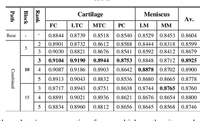
Abstract:Precise segmentation of knee tissues from magnetic resonance imaging (MRI) is critical in quantitative imaging and diagnosis. Convolutional neural networks (CNNs), which are state of the art, have limitations owing to the lack of image-specific adaptation, such as low tissue contrasts and structural inhomogeneities, thereby leading to incomplete segmentation results. This paper presents a deep learning based automatic segmentation framework for knee tissue segmentation. A novel multipath CNN-based method is proposed, which consists of an encoder decoder-based segmentation network in combination with a low rank tensor-reconstructed segmentation network. Low rank reconstruction in MRI tensor sub-blocks is introduced to exploit the structural and morphological variations in knee tissues. To further improve the segmentation from CNNs, trimap generation, which effectively utilizes superimposed regions, is proposed for defining high, medium and low confidence regions from the multipath CNNs. The secondary path with low rank reconstructed input mitigates the conditions in which the primary segmentation network can potentially fail and overlook the boundary regions. The outcome of the segmentation is solved as an alpha matting problem by blending the trimap with the source input. Experiments on Osteoarthritis Initiative (OAI) datasets and a self prepared scan validate the effectiveness of the proposed method. We specifically demonstrate the application of the proposed method in a cartilage segmentation based thickness map for diagnosis purposes.
Human Expression Recognition using Facial Shape Based Fourier Descriptors Fusion
Dec 28, 2020Abstract:Dynamic facial expression recognition has many useful applications in social networks, multimedia content analysis, security systems and others. This challenging process must be done under recurrent problems of image illumination and low resolution which changes at partial occlusions. This paper aims to produce a new facial expression recognition method based on the changes in the facial muscles. The geometric features are used to specify the facial regions i.e., mouth, eyes, and nose. The generic Fourier shape descriptor in conjunction with elliptic Fourier shape descriptor is used as an attribute to represent different emotions under frequency spectrum features. Afterwards a multi-class support vector machine is applied for classification of seven human expression. The statistical analysis showed our approach obtained overall competent recognition using 5-fold cross validation with high accuracy on well-known facial expression dataset.
 Add to Chrome
 Add to Firefox
 Add to Edge