Philipp Kainz
Semantic Segmentation of Colon Glands with Deep Convolutional Neural Networks and Total Variation Segmentation
Oct 10, 2017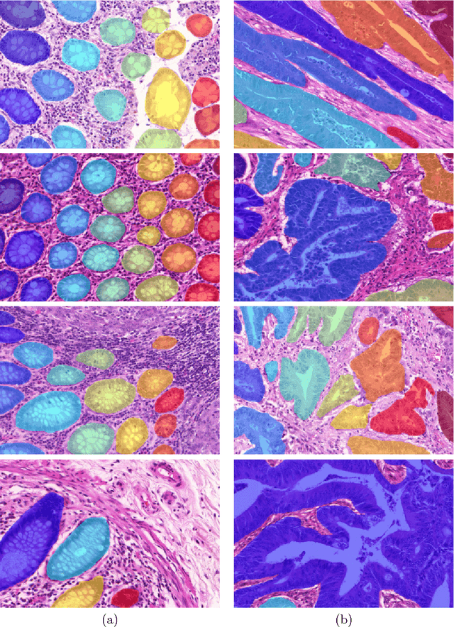
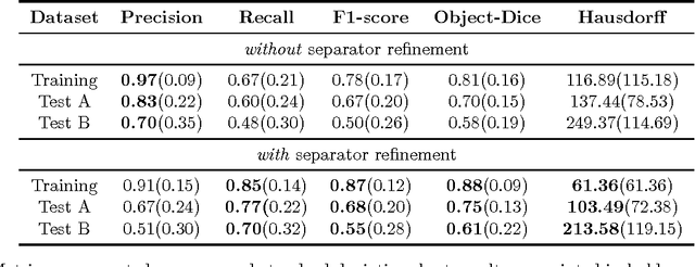
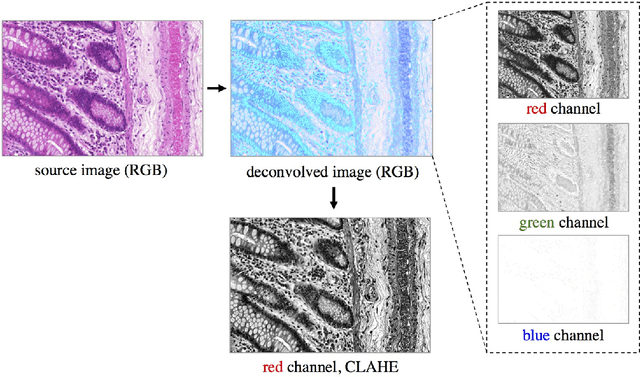
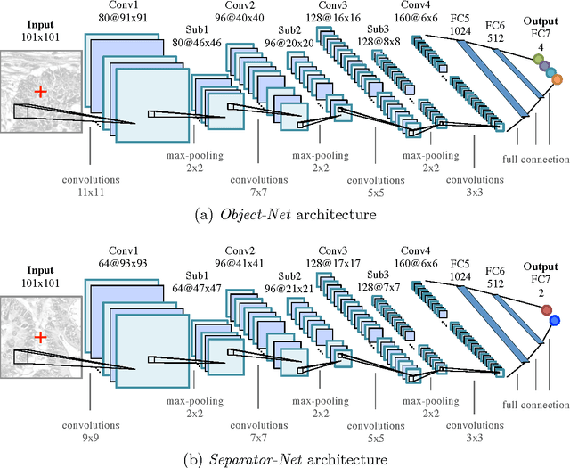
Abstract:Segmentation of histopathology sections is an ubiquitous requirement in digital pathology and due to the large variability of biological tissue, machine learning techniques have shown superior performance over standard image processing methods. As part of the GlaS@MICCAI2015 colon gland segmentation challenge, we present a learning-based algorithm to segment glands in tissue of benign and malignant colorectal cancer. Images are preprocessed according to the Hematoxylin-Eosin staining protocol and two deep convolutional neural networks (CNN) are trained as pixel classifiers. The CNN predictions are then regularized using a figure-ground segmentation based on weighted total variation to produce the final segmentation result. On two test sets, our approach achieves a tissue classification accuracy of 98% and 94%, making use of the inherent capability of our system to distinguish between benign and malignant tissue.
Gland Segmentation in Colon Histology Images: The GlaS Challenge Contest
Sep 01, 2016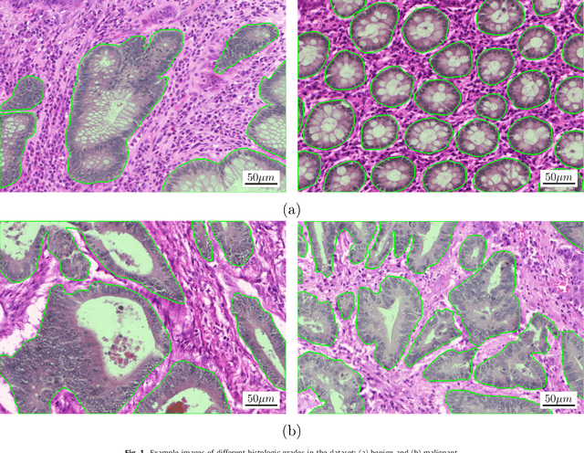
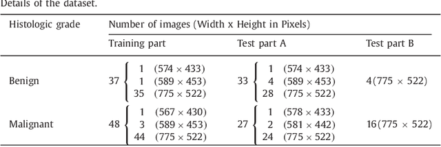
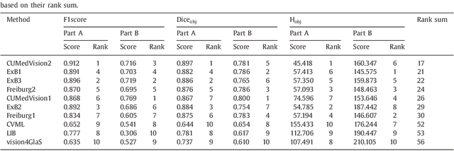
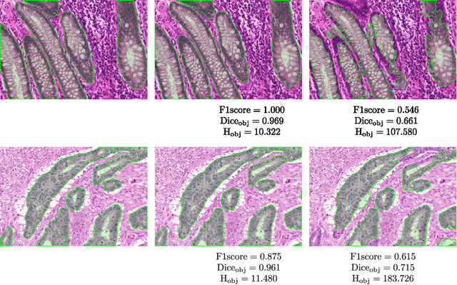
Abstract:Colorectal adenocarcinoma originating in intestinal glandular structures is the most common form of colon cancer. In clinical practice, the morphology of intestinal glands, including architectural appearance and glandular formation, is used by pathologists to inform prognosis and plan the treatment of individual patients. However, achieving good inter-observer as well as intra-observer reproducibility of cancer grading is still a major challenge in modern pathology. An automated approach which quantifies the morphology of glands is a solution to the problem. This paper provides an overview to the Gland Segmentation in Colon Histology Images Challenge Contest (GlaS) held at MICCAI'2015. Details of the challenge, including organization, dataset and evaluation criteria, are presented, along with the method descriptions and evaluation results from the top performing methods.
 Add to Chrome
 Add to Firefox
 Add to Edge