Philip Hoelter
Department of Neuroradiology, University Hospital Erlangen, Friedrich-Alexander-Universität
Noise and dose reduction in CT brain perfusion acquisition by projecting time attenuation curves onto lower dimensional spaces
Nov 02, 2021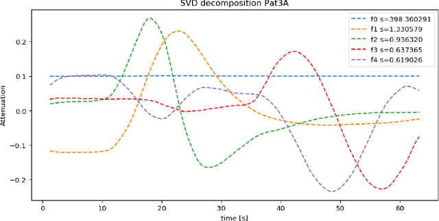

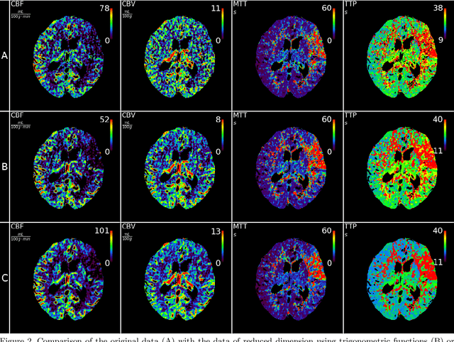

Abstract:CT perfusion imaging (CTP) plays an important role in decision making for the treatment of acute ischemic stroke with large vessel occlusion. Since the CT perfusion scan time is approximately one minute, the patient is exposed to a non-negligible dose of ionizing radiation. However, further dose reduction increases the level of noise in the data and the resulting perfusion maps. We present a method for reducing noise in perfusion data based on dimension reduction of time attenuation curves. For dimension reduction, we use either the fit of the first five terms of the trigonometric polynomial or the first five terms of the SVD decomposition of the time attenuation profiles. CTP data from four patients with large vessel occlusion and three control subjects were studied. To compare the noise level in the perfusion maps, we use the wavelet estimation of the noise standard deviation implemented in the scikit-image package. We show that both methods significantly reduce noise in the data while preserving important information about the perfusion deficits. These methods can be used to further reduce the dose in CT perfusion protocols or in perfusion studies using C-arm CT, which are burdened by high noise levels.
Time separation technique with the basis of trigonometric functions as an efficient method for flat detector CT brain perfusion imaging
Oct 18, 2021


Abstract:Dynamic perfusion imaging is routinely used in the diagnostic workup of acute ischemic stroke (AIS). At present, perfusion imaging can also be performed within the angio suite using flat detector computed tomography (FDCT). However, higher noise level, slower rotation speed and lower frame rate need to be considered in FDCT perfusion (FDCTP) data processing algorithms. The Time Separation Technique (TST) is a model-based perfusion data reconstruction method developed to solve these problems. In this contribution, we used TST and dimension reduction, where we approximate the time attenuation curves by a linear combination of trigonometric functions. Our goal was to show that TST with this data reduction does not impair clinical perfusion measurements. We performed a realistic simulation of FDCTP acquisition based on CT perfusion (CTP) data. Using these FDCTP data, we showed that TST provides better results than classical straightforward processing. Moreover we found that TST is robust to additional noise. Furthermore, we achieved a total processing time from reconstruction of FDCTP data to generation of perfusion maps of under 5 minutes. Perfusion maps created using TST with a trigonometric basis from FDCTP data show equivalent perfusion deficits as CT perfusion maps. Therefore, this technique can be considered a fast reliable tool for FDCTP imaging in AIS.
Temporal and Volumetric Denoising via Quantile Sparse Image (QuaSI) Prior in Optical Coherence Tomography and Beyond
Jul 05, 2018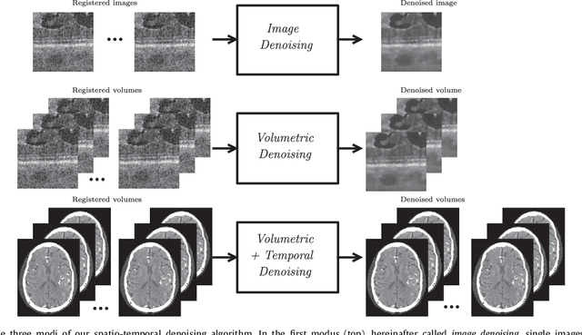

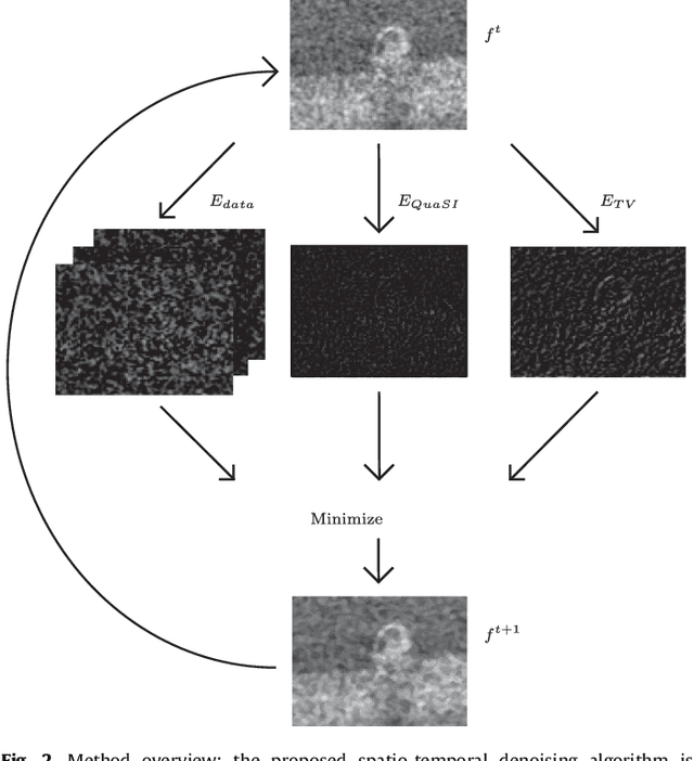
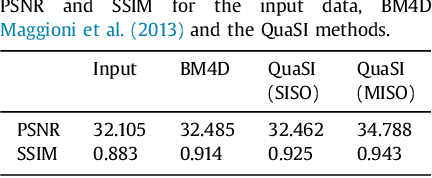
Abstract:This paper introduces an universal and structure-preserving regularization term, called quantile sparse image (QuaSI) prior. The prior is suitable for denoising images from various medical imaging modalities. We demonstrate its effectiveness on volumetric optical coherence tomography (OCT) and computed tomography (CT) data, which show different noise and image characteristics. OCT offers high-resolution scans of the human retina but is inherently impaired by speckle noise. CT on the other hand has a lower resolution and shows high-frequency noise. For the purpose of denoising, we propose a variational framework based on the QuaSI prior and a Huber data fidelity model that can handle 3-D and 3-D+t data. Efficient optimization is facilitated through the use of an alternating direction method of multipliers (ADMM) scheme and the linearization of the quantile filter. Experiments on multiple datasets emphasize the excellent performance of the proposed method.
 Add to Chrome
 Add to Firefox
 Add to Edge