 Add to Chrome
 Add to Firefox
 Add to Edge
EEG Interpretation Across Chant Listening: A Single-Subject Pilot Investigation Using Spectral and Functional Connectivity Analysis
Jun 23, 2026This technical report presents an EEG-based investigation of neural activity across five auditory conditions: Resting State (RS), Shiv Tandav Stotra (STS), Mahasudarshan Mantra (MM), Aum Chant, and Tanpura Listening. EEG recordings acquired from a healthy 5-year-old participant were analyzed using spectral power estimation and functional connectivity measures based on the weighted Phase Lag Index (wPLI). Spectral analysis revealed condition-specific modulation of neural oscillatory activity, with STS listening producing the highest relative power across multiple frequency bands, particularly within the beta range. Functional connectivity analysis demonstrated distinct network organizations across conditions. STS listening exhibited the strongest and most widespread connectivity pattern, characterized by prominent long-range interactions among frontal, temporal, parietal, and occipital regions. Tanpura listening generated a dense yet balanced connectivity network, while Aum listening showed moderate distributed connectivity. In contrast, MM and resting-state conditions displayed comparatively weaker and more localized network organization. These preliminary findings suggest that different chant-listening conditions engage distinct neural mechanisms involving both cortical activation and large-scale neural synchronization. The study establishes a methodological framework for future investigations examining the role of culturally relevant auditory interventions in cognitive development, neuroeducation, and child-centered neuroscience research.
Sex-based Network-Specific Differences in Connectomes: A Krakencoder-Based Analysis
Jun 15, 2026This study examines how deficiencies in one brain connectome modality propagate to the other, using the Krakencoder as a simulation framework. Structural and functional connectomes from 702 healthy participants in the Human Connectome Project were analyzed, with the impact of each of the Yeo-7 functional networks assessed separately. Seven scenarios were considered, each involving the removal of a single network while the remaining networks were preserved. The resulting perturbations in cross-modal predictions were quantified using three complementary metrics: KL divergence on eigenvalue spectra, Frobenius norm, and Wasserstein distance. In addition, the persistence of sex-specific information within the predicted connectomes was evaluated. Across all metrics and both prediction directions, the Default Mode Network produced the largest perturbations, whereas the Somatomotor network yielded the smallest. Sex differences in network-level perturbation signatures were subtle, with the best result being an accuracy of 66.09% from connectomes predicted under network-removal conditions. In contrast, connectomes predicted from intact inputs achieved substantially higher sex classification accuracy, reaching up to 84.76%. These findings confirm that full predicted connectomes retain considerably more sex-discriminative information than perturbation-derived signatures alone.
Hybrid Classical-Quantum (HCQ) Alzheimer's Classification via Supervised $β$-VAE and Quantum Kernels
Jun 12, 2026This paper presents a two-stage Hybrid Classical-Quantum (HCQ) pipeline for binary Alzheimer's disease (AD) classification from 3D T1-weighted structural MRI volumes, where the classical and quantum components are designed to complement each other rather than operate independently. A supervised 3D $β$-variational autoencoder (VAE) is trained end-to-end under voxel-wise reconstruction, KL-divergence, and focal classification losses that compress each 3D MRI volume (resized from 152 x 184 x 152 to 96 x 96 x 96) into a 64-dimensional latent code. Partial Least Squares (PLS) regression selects the six components in the latent code that best separate Alzheimer's Disease (AD) from cognitively normal (CN) subjects and rescales them into rotation angles, which are encoded onto a six-qubit register using the ZZ quantum feature map to give us the respective quantum states. The input to a precomputed-kernel Support Vector Machine (SVM) is an N x N Gram matrix (N = 308), created by calculating the overlap between every pair of quantum states. The novelty of this work lies in the fact that the quantum kernel operates directly on disease-aware features that are learned end-to-end by a supervised autoencoder, rather than on pre-extracted inputs. On 308 ADNI-1 subjects, consisting of 137 AD and 171 CN subjects, the baseline achieved 67.2% accuracy and 0.759 AUC, while the stability-enhanced variant reached 72.1% accuracy and 0.799 AUC with cross-fold variance halved. 3D Grad-CAM further helped validate our model's focus on brain regions linked to Alzheimer's. The HCQ pipeline could serve as a general-purpose framework for diagnostic classification across biomedical imaging domains that present similar challenges for classical approaches.
Multi-Task Learning with Additive U-Net for Image Denoising and Classification
Feb 13, 2026We investigate additive skip fusion in U-Net architectures for image denoising and denoising-centric multi-task learning (MTL). By replacing concatenative skips with gated additive fusion, the proposed Additive U-Net (AddUNet) constrains shortcut capacity while preserving fixed feature dimensionality across depth. This structural regularization induces controlled encoder-decoder information flow and stabilizes joint optimization. Across single-task denoising and joint denoising-classification settings, AddUNet achieves competitive reconstruction performance with improved training stability. In MTL, learned skip weights exhibit systematic task-aware redistribution: shallow skips favor reconstruction, while deeper features support discrimination. Notably, reconstruction remains robust even under limited classification capacity, indicating implicit task decoupling through additive fusion. These findings show that simple constraints on skip connections act as an effective architectural regularizer for stable and scalable multi-task learning without increasing model complexity.
Chest X-ray Classification using Deep Convolution Models on Low-resolution images with Uncertain Labels
Apr 12, 2025Deep Convolutional Neural Networks have consistently proven to achieve state-of-the-art results on a lot of imaging tasks over the past years' majority of which comprise of high-quality data. However, it is important to work on low-resolution images since it could be a cheaper alternative for remote healthcare access where the primary need of automated pathology identification models occurs. Medical diagnosis using low-resolution images is challenging since critical details may not be easily identifiable. In this paper, we report classification results by experimenting on different input image sizes of Chest X-rays to deep CNN models and discuss the feasibility of classification on varying image sizes. We also leverage the noisy labels in the dataset by proposing a Randomized Flipping of labels techniques. We use an ensemble of multi-label classification models on frontal and lateral studies. Our models are trained on 5 out of the 14 chest pathologies of the publicly available CheXpert dataset. We incorporate techniques such as augmentation, regularization for model improvement and use class activation maps to visualize the neural network's decision making. Comparison with classification results on data from 200 subjects, obtained on the corresponding high-resolution images, reported in the original CheXpert paper, has been presented. For pathologies Cardiomegaly, Consolidation and Edema, we obtain 3% higher accuracy with our model architecture.
Graph Classification and Radiomics Signature for Identification of Tuberculous Meningitis
Apr 01, 2025
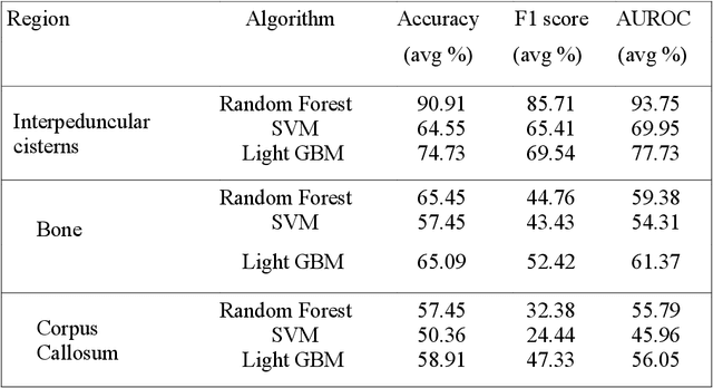


Introduction: Tuberculous meningitis (TBM) is a serious brain infection caused by Mycobacterium tuberculosis, characterized by inflammation of the meninges covering the brain and spinal cord. Diagnosis often requires invasive lumbar puncture (LP) and cerebrospinal fluid (CSF) analysis. Objectives: This study aims to classify TBM patients using T1-weighted (T1w) non-contrast Magnetic Resonance Imaging (MRI) scans. We hypothesize that specific brain regions, such as the interpeduncular cisterns, bone, and corpus callosum, contain visual markers that can non-invasively distinguish TBM patients from healthy controls. We propose a novel Pixel-array Graphs Classifier (PAG-Classifier) that leverages spatial relationships between neighbouring 3D pixels in a graph-based framework to extract significant features through eigen decomposition. These features are then used to train machine learning classifiers for effective patient classification. We validate our approach using a radiomics-based methodology, classifying TBM patients based on relevant radiomics features. Results: We utilized an internal dataset consisting of 52 scans, 32 from confirmed TBM patients based on mycobacteria detection in CSF, and 20 from healthy individuals. We achieved a 5-fold cross-validated average F1 score of 85.71% for cistern regions with our PAG-Classifier and 92.85% with the radiomics features classifier, surpassing current state-of-the-art benchmarks by 15% and 22%, respectively. However, bone and corpus callosum regions showed poor classification effectiveness, with average F1 scores below 50%. Conclusion: Our study suggests that algorithms like the PAG-Classifier serve as effective tools for non-invasive TBM analysis, particularly by targeting the interpeduncular cistern. Findings indicate that the bone and corpus callosum regions lack distinctive patterns for differentiation.
Alzheimer's Disease Classification Using Retinal OCT: TransnetOCT and Swin Transformer Models
Mar 14, 2025Retinal optical coherence tomography (OCT) images are the biomarkers for neurodegenerative diseases, which are rising in prevalence. Early detection of Alzheimer's disease using retinal OCT is a primary challenging task. This work utilizes advanced deep learning techniques to classify retinal OCT images of subjects with Alzheimer's disease (AD) and healthy controls (CO). The goal is to enhance diagnostic capabilities through efficient image analysis. In the proposed model, Raw OCT images have been preprocessed with ImageJ and given to various deep-learning models to evaluate the accuracy. The best classification architecture is TransNetOCT, which has an average accuracy of 98.18% for input OCT images and 98.91% for segmented OCT images for five-fold cross-validation compared to other models, and the Swin Transformer model has achieved an accuracy of 93.54%. The evaluation accuracy metric demonstrated TransNetOCT and Swin transformer models capability to classify AD and CO subjects reliably, contributing to the potential for improved diagnostic processes in clinical settings.
Persistent Homology for MCI Classification: A Comparative Analysis between Graph and Vietoris-Rips Filtrations
Oct 30, 2024


Mild cognitive impairment (MCI), often linked to early neurodegeneration, is characterized by subtle cognitive declines and disruptions in brain connectivity. The present study offers a detailed analysis of topological changes associated with MCI, focusing on two subtypes: Early MCI and Late MCI. This analysis utilizes fMRI time series data from two distinct populations: the publicly available ADNI dataset (Western cohort) and the in-house TLSA dataset (Indian Urban cohort). Persistent Homology, a topological data analysis method, is employed with two distinct filtration techniques - Vietoris-Rips and graph filtration-for classifying MCI subtypes. For Vietoris-Rips filtration, inter-ROI Wasserstein distance matrices between persistent diagrams are used for classification, while graph filtration relies on the top ten most persistent homology features. Comparative analysis shows that the Vietoris-Rips filtration significantly outperforms graph filtration, capturing subtle variations in brain connectivity with greater accuracy. The Vietoris-Rips filtration method achieved the highest classification accuracy of 85.7\% for distinguishing between age and gender matched healthy controls and MCI, whereas graph filtration reached a maximum accuracy of 71.4\% for the same task. This superior performance highlights the sensitivity of Vietoris-Rips filtration in detecting intricate topological features associated with neurodegeneration. The findings underscore the potential of persistent homology, particularly when combined with the Wasserstein distance, as a powerful tool for early diagnosis and precise classification of cognitive impairments, offering valuable insights into brain connectivity changes in MCI.
Leveraging Persistent Homology for Differential Diagnosis of Mild Cognitive Impairment
Aug 28, 2024


Mild cognitive impairment (MCI) is characterized by subtle changes in cognitive functions, often associated with disruptions in brain connectivity. The present study introduces a novel fine-grained analysis to examine topological alterations in neurodegeneration pertaining to six different brain networks of MCI subjects (Early/Late MCI). To achieve this, fMRI time series from two distinct populations are investigated: (i) the publicly accessible ADNI dataset and (ii) our in-house dataset. The study utilizes sliding window embedding to convert each fMRI time series into a sequence of 3-dimensional vectors, facilitating the assessment of changes in regional brain topology. Distinct persistence diagrams are computed for Betti descriptors of dimension-0, 1, and 2. Wasserstein distance metric is used to quantify differences in topological characteristics. We have examined both (i) ROI-specific inter-subject interactions and (ii) subject-specific inter-ROI interactions. Further, a new deep learning model is proposed for classification, achieving a maximum classification accuracy of 95% for the ADNI dataset and 85% for the in-house dataset. This methodology is further adapted for the differential diagnosis of MCI sub-types, resulting in a peak accuracy of 76.5%, 91.1% and 80% in classifying HC Vs. EMCI, HC Vs. LMCI and EMCI Vs. LMCI, respectively. We showed that the proposed approach surpasses current state-of-the-art techniques designed for classifying MCI and its sub-types using fMRI.
Non-linear Analysis Based ECG Classification of Cardiovascular Disorders
Aug 02, 2024


Multi-channel ECG-based cardiac disorders detection has an impact on cardiac care and treatment. Limitations of existing methods included variation in ECG waveforms due to the location of electrodes, high non-linearity in the signal, and amplitude measurement in millivolts. The present study reports a non-linear analysis-based methodology that utilizes Recurrence plot visualization. The patterned occurrence of well-defined structures, such as the QRS complex, can be exploited effectively using Recurrence plots. This Recurrence-based method is applied to the publicly available Physikalisch-Technische Bundesanstalt (PTB) dataset from PhysioNet database, where we studied four classes of different cardiac disorders (Myocardial infarction, Bundle branch blocks, Cardiomyopathy, and Dysrhythmia) and healthy controls, achieving an impressive classification accuracy of 100%. Additionally, t-SNE plot visualizations of the latent space embeddings derived from Recurrence plots and Recurrence Quantification Analysis features reveal a clear demarcation between the considered cardiac disorders and healthy individuals, demonstrating the potential of this approach.