Muhammad Abdul Kadir
Multimodal EEG and Keystroke Dynamics Based Biometric System Using Machine Learning Algorithms
Mar 10, 2021


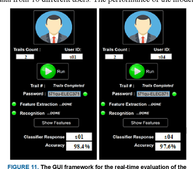
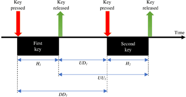
Abstract:With the rapid advancement of technology, different biometric user authentication, and identification systems are emerging. Traditional biometric systems like face, fingerprint, and iris recognition, keystroke dynamics, etc. are prone to cyber-attacks and suffer from different disadvantages. Electroencephalography (EEG) based authentication has shown promise in overcoming these limitations. However, EEG-based authentication is less accurate due to signal variability at different psychological and physiological conditions. On the other hand, keystroke dynamics-based identification offers high accuracy but suffers from different spoofing attacks. To overcome these challenges, we propose a novel multimodal biometric system combining EEG and keystroke dynamics. Firstly, a dataset was created by acquiring both keystroke dynamics and EEG signals from 10 users with 500 trials per user at 10 different sessions. Different statistical, time, and frequency domain features were extracted and ranked from the EEG signals and key features were extracted from the keystroke dynamics. Different classifiers were trained, validated, and tested for both individual and combined modalities for two different classification strategies - personalized and generalized. Results show that very high accuracy can be achieved both in generalized and personalized cases for the combination of EEG and keystroke dynamics. The identification and authentication accuracies were found to be 99.80% and 99.68% for Extreme Gradient Boosting (XGBoost) and Random Forest classifiers, respectively which outperform the individual modalities with a significant margin (around 5 percent). We also developed a binary template matching-based algorithm, which gives 93.64% accuracy 6X faster. The proposed method is secured and reliable for any kind of biometric authentication.
Reliable Tuberculosis Detection using Chest X-ray with Deep Learning, Segmentation and Visualization
Jul 29, 2020


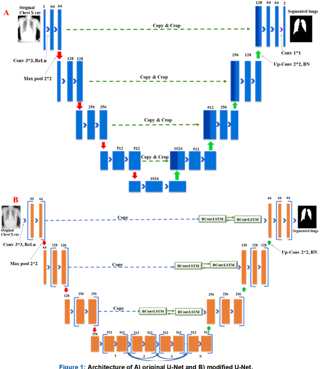
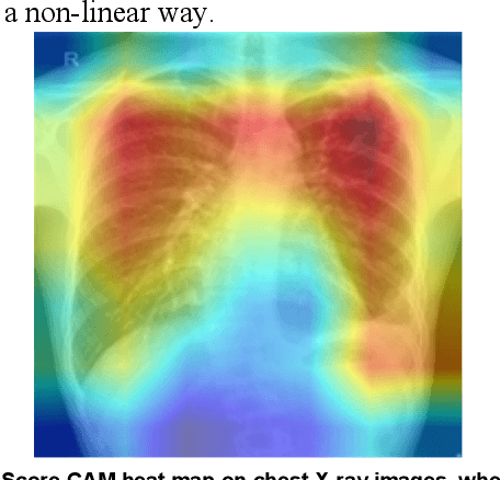
Abstract:Tuberculosis (TB) is a chronic lung disease that occurs due to bacterial infection and is one of the top 10 leading causes of death. Accurate and early detection of TB is very important, otherwise, it could be life-threatening. In this work, we have detected TB reliably from the chest X-ray images using image pre-processing, data augmentation, image segmentation, and deep-learning classification techniques. Several public databases were used to create a database of 700 TB infected and 3500 normal chest X-ray images for this study. Nine different deep CNNs (ResNet18, ResNet50, ResNet101, ChexNet, InceptionV3, Vgg19, DenseNet201, SqueezeNet, and MobileNet), which were used for transfer learning from their pre-trained initial weights and trained, validated and tested for classifying TB and non-TB normal cases. Three different experiments were carried out in this work: segmentation of X-ray images using two different U-net models, classification using X-ray images, and segmented lung images. The accuracy, precision, sensitivity, F1-score, specificity in the detection of tuberculosis using X-ray images were 97.07 %, 97.34 %, 97.07 %, 97.14 % and 97.36 % respectively. However, segmented lungs for the classification outperformed than whole X-ray image-based classification and accuracy, precision, sensitivity, F1-score, specificity were 99.9 %, 99.91 %, 99.9 %, 99.9 %, and 99.52 % respectively. The paper also used a visualization technique to confirm that CNN learns dominantly from the segmented lung regions results in higher detection accuracy. The proposed method with state-of-the-art performance can be useful in the computer-aided faster diagnosis of tuberculosis.
Can AI help in screening Viral and COVID-19 pneumonia?
Apr 28, 2020

Abstract:Coronavirus disease (COVID-19) is a pandemic disease, which has already infected around 3 million people and caused fatalities of above 200 thousand. The aim of this paper is to automatically detect COVID-19 pneumonia patients using digital x-ray images while maximizing the accuracy in detection using image pre-processing and deep-learning techniques. A public database was created by the authors using three public databases and by collecting images from recently published articles. The database contains a mixture of 190 COVID-19, 1345 viral pneumonia, and 1341 normal chest x-ray images. An image augmented training set was created with around 2600 images of each category for training and validating four different pre-trained deep Convolutional Neural Networks (CNNs). These networks were tested for the classification of two different schemes (normal and COVID-19 pneumonia; normal, viral and COVID-19 pneumonia). The classification accuracy, sensitivity, specificity and precision for both the schemes were 98.3%, 96.7%, 100%, 100% and 98.3%, 96.7%, 99%, 100%, respectively. The high accuracy of this computer-aided diagnostic tool can significantly improve the speed and accuracy of diagnosing cases with COVID-19. This would be highly useful in this pandemic where disease burden and need for preventive measures are at odds with available resources.
 Add to Chrome
 Add to Firefox
 Add to Edge