Maxim Kan
Towards Unpaired Depth Enhancement and Super-Resolution in the Wild
May 25, 2021
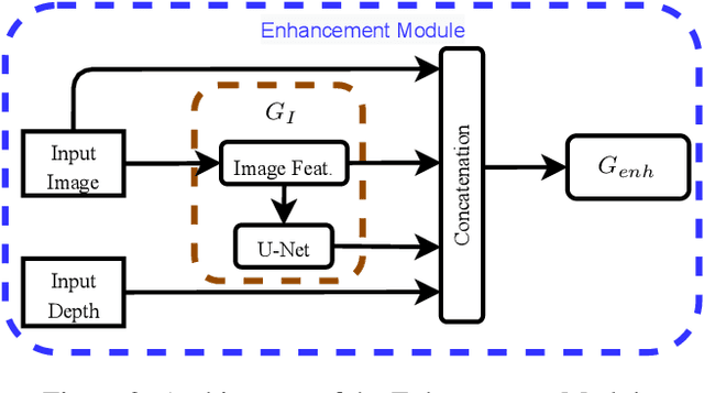

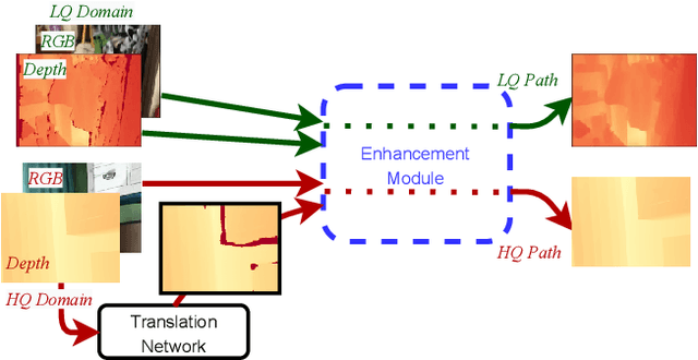
Abstract:Depth maps captured with commodity sensors are often of low quality and resolution; these maps need to be enhanced to be used in many applications. State-of-the-art data-driven methods of depth map super-resolution rely on registered pairs of low- and high-resolution depth maps of the same scenes. Acquisition of real-world paired data requires specialized setups. Another alternative, generating low-resolution maps from high-resolution maps by subsampling, adding noise and other artificial degradation methods, does not fully capture the characteristics of real-world low-resolution images. As a consequence, supervised learning methods trained on such artificial paired data may not perform well on real-world low-resolution inputs. We consider an approach to depth map enhancement based on learning from unpaired data. While many techniques for unpaired image-to-image translation have been proposed, most are not directly applicable to depth maps. We propose an unpaired learning method for simultaneous depth enhancement and super-resolution, which is based on a learnable degradation model and surface normal estimates as features to produce more accurate depth maps. We demonstrate that our method outperforms existing unpaired methods and performs on par with paired methods on a new benchmark for unpaired learning that we developed.
Interpretable Deep Learning for Pattern Recognition in Brain Differences Between Men and Women
Jun 20, 2020

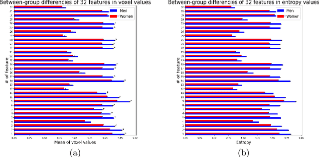
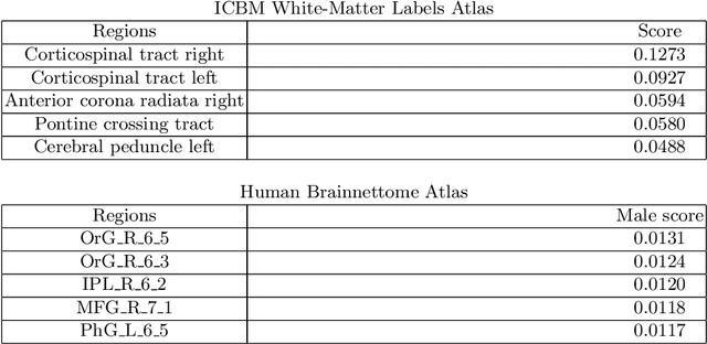
Abstract:Deep learning shows high potential for many medical image analysis tasks. Neural networks work with full-size data without extensive preprocessing and feature generation and, thus, information loss. Recent work has shown that morphological difference between specific brain regions can be found on MRI with deep learning techniques. We consider the pattern recognition task based on a large open-access dataset of healthy subjects - an exploration of brain differences between men and women. However, interpretation of the lately proposed models is based on a region of interest and can not be extended to pixel or voxel-wise image interpretation, which is considered to be more informative. In this paper, we confirm the previous findings in sex differences from diffusion-tensor imaging on T1 weighted brain MRI scans. We compare the results of three voxel-based 3D CNN interpretation methods: Meaningful Perturbations, GradCam and Guided Backpropagation and provide the open-source code.
 Add to Chrome
 Add to Firefox
 Add to Edge