Lorenzo Preda
Comparative clinical evaluation of "memory-efficient" synthetic 3d generative adversarial networks (gan) head-to-head to state of art: results on computed tomography of the chest
Jan 26, 2025Abstract:Introduction: Generative Adversarial Networks (GANs) are increasingly used to generate synthetic medical images, addressing the critical shortage of annotated data for training Artificial Intelligence (AI) systems. This study introduces a novel memory-efficient GAN architecture, incorporating Conditional Random Fields (CRFs) to generate high-resolution 3D medical images and evaluates its performance against the state-of-the-art hierarchical (HA)-GAN model. Materials and Methods: The CRF-GAN was trained using the open-source lung CT LUNA16 dataset. The architecture was compared to HA-GAN through a quantitative evaluation, using Frechet Inception Distance (FID) and Maximum Mean Discrepancy (MMD) metrics, and a qualitative evaluation, through a two-alternative forced choice (2AFC) test completed by a pool of 12 resident radiologists, in order to assess the realism of the generated images. Results: CRF-GAN outperformed HA-GAN with lower FID (0.047 vs. 0.061) and MMD (0.084 vs. 0.086) scores, indicating better image fidelity. The 2AFC test showed a significant preference for images generated by CRF-Gan over those generated by HA-GAN with a p-value of 1.93e-05. Additionally, CRF-GAN demonstrated 9.34% lower memory usage at 256 resolution and achieved up to 14.6% faster training speeds, offering substantial computational savings. Discussion: CRF-GAN model successfully generates high-resolution 3D medical images with non-inferior quality to conventional models, while being more memory-efficient and faster. Computational power and time saved can be used to improve the spatial resolution and anatomical accuracy of generated images, which is still a critical factor limiting their direct clinical applicability.
Reshaping Free-Text Radiology Notes Into Structured Reports With Generative Transformers
Mar 27, 2024Abstract:BACKGROUND: Radiology reports are typically written in a free-text format, making clinical information difficult to extract and use. Recently the adoption of structured reporting (SR) has been recommended by various medical societies thanks to the advantages it offers, e.g. standardization, completeness and information retrieval. We propose a pipeline to extract information from free-text radiology reports, that fits with the items of the reference SR registry proposed by a national society of interventional and medical radiology, focusing on CT staging of patients with lymphoma. METHODS: Our work aims to leverage the potential of Natural Language Processing (NLP) and Transformer-based models to deal with automatic SR registry filling. With the availability of 174 radiology reports, we investigate a rule-free generative Question Answering approach based on a domain-specific version of T5 (IT5). Two strategies (batch-truncation and ex-post combination) are implemented to comply with the model's context length limitations. Performance is evaluated in terms of strict accuracy, F1, and format accuracy, and compared with the widely used GPT-3.5 Large Language Model. A 5-point Likert scale questionnaire is used to collect human-expert feedback on the similarity between medical annotations and generated answers. RESULTS: The combination of fine-tuning and batch splitting allows IT5 to achieve notable results; it performs on par with GPT-3.5 albeit its size being a thousand times smaller in terms of parameters. Human-based assessment scores show a high correlation (Spearman's correlation coefficients>0.88, p-values<0.001) with AI performance metrics (F1) and confirm the superior ability of LLMs (i.e., GPT-3.5, 175B of parameters) in generating plausible human-like statements.
AIforCOVID: predicting the clinical outcomes in patients with COVID-19 applying AI to chest-X-rays. An Italian multicentre study
Dec 11, 2020


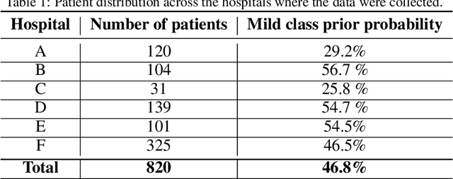
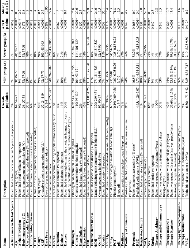
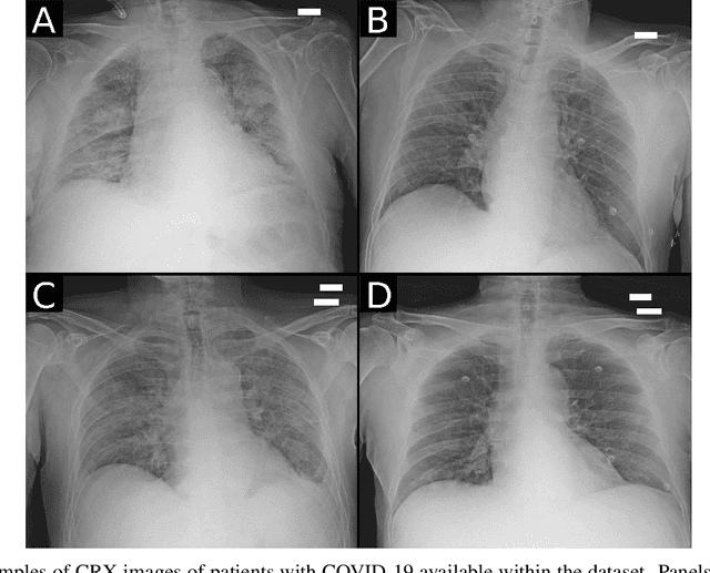
Abstract:Recent epidemiological data report that worldwide more than 53 million people have been infected by SARS-CoV-2, resulting in 1.3 million deaths. The disease has been spreading very rapidly and few months after the identification of the first infected, shortage of hospital resources quickly became a problem. In this work we investigate whether chest X-ray (CXR) can be used as a possible tool for the early identification of patients at risk of severe outcome, like intensive care or death. CXR is a radiological technique that compared to computed tomography (CT) it is simpler, faster, more widespread and it induces lower radiation dose. We present a dataset including data collected from 820 patients by six Italian hospitals in spring 2020 during the first COVID-19 emergency. The dataset includes CXR images, several clinical attributes and clinical outcomes. We investigate the potential of artificial intelligence to predict the prognosis of such patients, distinguishing between severe and mild cases, thus offering a baseline reference for other researchers and practitioners. To this goal, we present three approaches that use features extracted from CXR images, either handcrafted or automatically by convolutional neuronal networks, which are then integrated with the clinical data. Exhaustive evaluation shows promising performance both in 10-fold and leave-one-centre-out cross-validation, implying that clinical data and images have the potential to provide useful information for the management of patients and hospital resources.
 Add to Chrome
 Add to Firefox
 Add to Edge