Giovanni Valbusa
LIP-CAR: contrast agent reduction by a deep learned inverse problem
Jul 15, 2024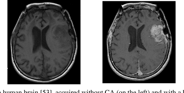
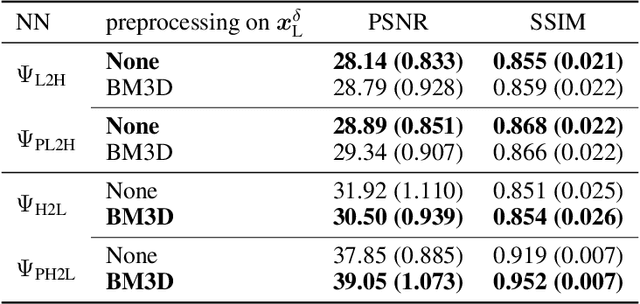
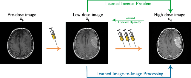

Abstract:The adoption of contrast agents in medical imaging protocols is crucial for accurate and timely diagnosis. While highly effective and characterized by an excellent safety profile, the use of contrast agents has its limitation, including rare risk of allergic reactions, potential environmental impact and economic burdens on patients and healthcare systems. In this work, we address the contrast agent reduction (CAR) problem, which involves reducing the administered dosage of contrast agent while preserving the visual enhancement. The current literature on the CAR task is based on deep learning techniques within a fully image processing framework. These techniques digitally simulate high-dose images from images acquired with a low dose of contrast agent. We investigate the feasibility of a ``learned inverse problem'' (LIP) approach, as opposed to the end-to-end paradigm in the state-of-the-art literature. Specifically, we learn the image-to-image operator that maps high-dose images to their corresponding low-dose counterparts, and we frame the CAR task as an inverse problem. We then solve this problem through a regularized optimization reformulation. Regularization methods are well-established mathematical techniques that offer robustness and explainability. Our approach combines these rigorous techniques with cutting-edge deep learning tools. Numerical experiments performed on pre-clinical medical images confirm the effectiveness of this strategy, showing improved stability and accuracy in the simulated high-dose images.
AIforCOVID: predicting the clinical outcomes in patients with COVID-19 applying AI to chest-X-rays. An Italian multicentre study
Dec 11, 2020
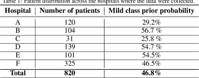
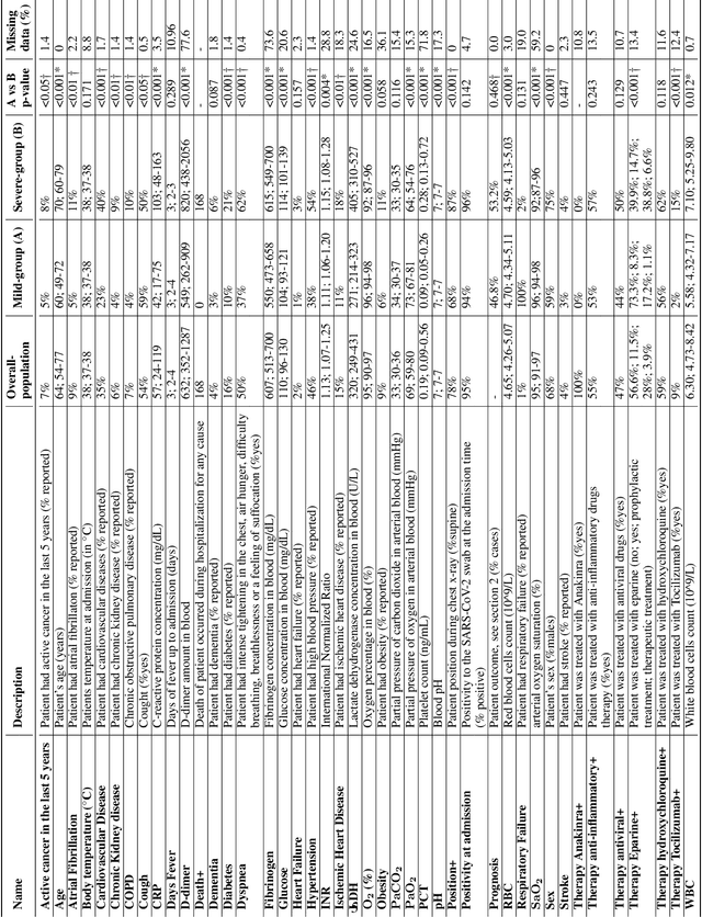
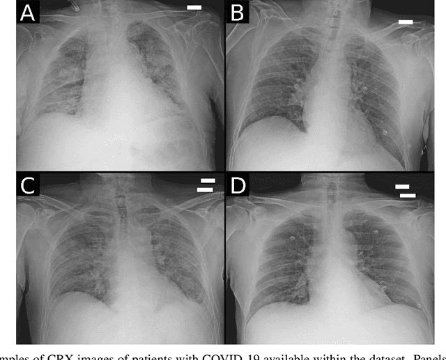
Abstract:Recent epidemiological data report that worldwide more than 53 million people have been infected by SARS-CoV-2, resulting in 1.3 million deaths. The disease has been spreading very rapidly and few months after the identification of the first infected, shortage of hospital resources quickly became a problem. In this work we investigate whether chest X-ray (CXR) can be used as a possible tool for the early identification of patients at risk of severe outcome, like intensive care or death. CXR is a radiological technique that compared to computed tomography (CT) it is simpler, faster, more widespread and it induces lower radiation dose. We present a dataset including data collected from 820 patients by six Italian hospitals in spring 2020 during the first COVID-19 emergency. The dataset includes CXR images, several clinical attributes and clinical outcomes. We investigate the potential of artificial intelligence to predict the prognosis of such patients, distinguishing between severe and mild cases, thus offering a baseline reference for other researchers and practitioners. To this goal, we present three approaches that use features extracted from CXR images, either handcrafted or automatically by convolutional neuronal networks, which are then integrated with the clinical data. Exhaustive evaluation shows promising performance both in 10-fold and leave-one-centre-out cross-validation, implying that clinical data and images have the potential to provide useful information for the management of patients and hospital resources.
 Add to Chrome
 Add to Firefox
 Add to Edge