Kevin G. Field
Accelerating Domain-Aware Electron Microscopy Analysis Using Deep Learning Models with Synthetic Data and Image-Wide Confidence Scoring
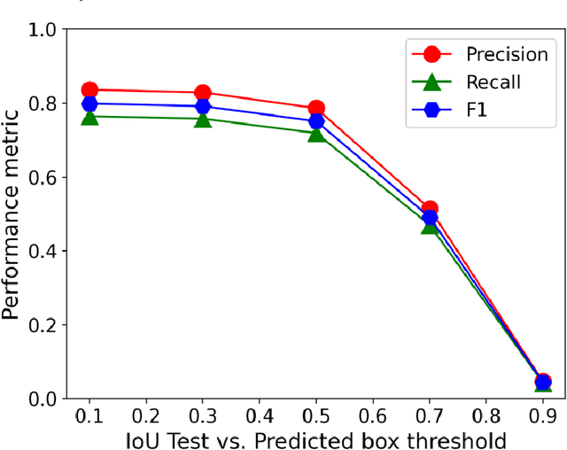
Aug 02, 2024Abstract:The integration of machine learning (ML) models enhances the efficiency, affordability, and reliability of feature detection in microscopy, yet their development and applicability are hindered by the dependency on scarce and often flawed manually labeled datasets and a lack of domain awareness. We addressed these challenges by creating a physics-based synthetic image and data generator, resulting in a machine learning model that achieves comparable precision (0.86), recall (0.63), F1 scores (0.71), and engineering property predictions (R2=0.82) to a model trained on human-labeled data. We enhanced both models by using feature prediction confidence scores to derive an image-wide confidence metric, enabling simple thresholding to eliminate ambiguous and out-of-domain images resulting in performance boosts of 5-30% with a filtering-out rate of 25%. Our study demonstrates that synthetic data can eliminate human reliance in ML and provides a means for domain awareness in cases where many feature detections per image are needed.
Performance, Successes and Limitations of Deep Learning Semantic Segmentation of Multiple Defects in Transmission Electron Micrographs
Oct 15, 2021


Abstract:In this work, we perform semantic segmentation of multiple defect types in electron microscopy images of irradiated FeCrAl alloys using a deep learning Mask Regional Convolutional Neural Network (Mask R-CNN) model. We conduct an in-depth analysis of key model performance statistics, with a focus on quantities such as predicted distributions of defect shapes, defect sizes, and defect areal densities relevant to informing modeling and understanding of irradiated Fe-based materials properties. To better understand the performance and present limitations of the model, we provide examples of useful evaluation tests which include a suite of random splits, and dataset size-dependent and domain-targeted cross validation tests. Overall, we find that the current model is a fast, effective tool for automatically characterizing and quantifying multiple defect types in microscopy images, with a level of accuracy on par with human domain expert labelers. More specifically, the model can achieve average defect identification F1 scores as high as 0.8, and, based on random cross validation, have low overall average (+/- standard deviation) defect size and density percentage errors of 7.3 (+/- 3.8)% and 12.7 (+/- 5.3)%, respectively. Further, our model predicts the expected material hardening to within 10-20 MPa (about 10% of total hardening), which is about the same error level as experiments. Our targeted evaluation tests also suggest the best path toward improving future models is not expanding existing databases with more labeled images but instead data additions that target weak points of the model domain, such as images from different microscopes, imaging conditions, irradiation environments, and alloy types. Finally, we discuss the first phase of an effort to provide an easy-to-use, open-source object detection tool to the broader community for identifying defects in new images.
A Deep Learning Based Automatic Defect Analysis Framework for In-situ TEM Ion Irradiations
Aug 19, 2021


Abstract:Videos captured using Transmission Electron Microscopy (TEM) can encode details regarding the morphological and temporal evolution of a material by taking snapshots of the microstructure sequentially. However, manual analysis of such video is tedious, error-prone, unreliable, and prohibitively time-consuming if one wishes to analyze a significant fraction of frames for even videos of modest length. In this work, we developed an automated TEM video analysis system for microstructural features based on the advanced object detection model called YOLO and tested the system on an in-situ ion irradiation TEM video of dislocation loops formed in a FeCrAl alloy. The system provides analysis of features observed in TEM including both static and dynamic properties using the YOLO-based defect detection module coupled to a geometry analysis module and a dynamic tracking module. Results show that the system can achieve human comparable performance with an F1 score of 0.89 for fast, consistent, and scalable frame-level defect analysis. This result is obtained on a real but exceptionally clean and stable data set and more challenging data sets may not achieve this performance. The dynamic tracking also enabled evaluation of individual defect evolution like per defect growth rate at a fidelity never before achieved using common human analysis methods. Our work shows that automatically detecting and tracking interesting microstructures and properties contained in TEM videos is viable and opens new doors for evaluating materials dynamics.
 Add to Chrome
 Add to Firefox
 Add to Edge