Guillaume Dardenne
Suicide Risk Assessment Using Multimodal Speech Features: A Study on the SW1 Challenge Dataset
May 19, 2025Abstract:The 1st SpeechWellness Challenge conveys the need for speech-based suicide risk assessment in adolescents. This study investigates a multimodal approach for this challenge, integrating automatic transcription with WhisperX, linguistic embeddings from Chinese RoBERTa, and audio embeddings from WavLM. Additionally, handcrafted acoustic features -- including MFCCs, spectral contrast, and pitch-related statistics -- were incorporated. We explored three fusion strategies: early concatenation, modality-specific processing, and weighted attention with mixup regularization. Results show that weighted attention provided the best generalization, achieving 69% accuracy on the development set, though a performance gap between development and test sets highlights generalization challenges. Our findings, strictly tied to the MINI-KID framework, emphasize the importance of refining embedding representations and fusion mechanisms to enhance classification reliability.
Fully automated workflow for the design of patient-specific orthopaedic implants: application to total knee arthroplasty
Mar 25, 2024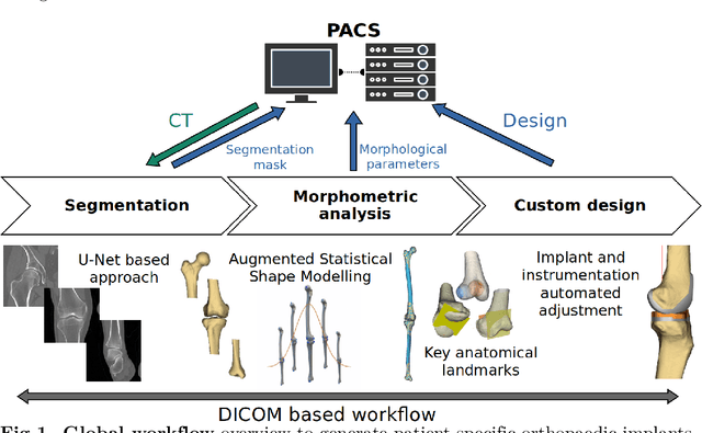

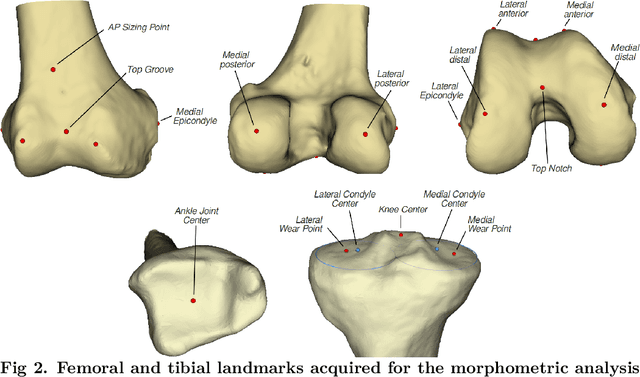
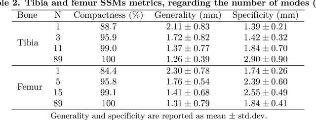
Abstract:Arthroplasty is commonly performed to treat joint osteoarthritis, reducing pain and improving mobility. While arthroplasty has known several technical improvements, a significant share of patients are still unsatisfied with their surgery. Personalised arthroplasty improves surgical outcomes however current solutions require delays, making it difficult to integrate in clinical routine. We propose a fully automated workflow to design patient-specific implants, presented for total knee arthroplasty, the most widely performed arthroplasty in the world nowadays. The proposed pipeline first uses artificial neural networks to segment the proximal and distal extremities of the femur and tibia. Then the full bones are reconstructed using augmented statistical shape models, combining shape and landmarks information. Finally, 77 morphological parameters are computed to design patient-specific implants. The developed workflow has been trained using 91 CT scans of lower limb and evaluated on 41 CT scans manually segmented, in terms of accuracy and execution time. The workflow accuracy was $0.4\pm0.2mm$ for the segmentation, $1.2\pm0.4mm$ for the full bones reconstruction, and $2.8\pm2.2mm$ for the anatomical landmarks determination. The custom implants fitted the patients' anatomy with $0.6\pm0.2mm$ accuracy. The whole process from segmentation to implants' design lasted about 5 minutes. The proposed workflow allows for a fast and reliable personalisation of knee implants, directly from the patient CT image without requiring any manual intervention. It establishes a patient-specific pre-operative planning for TKA in a very short time making it easily available for all patients. Combined with efficient implant manufacturing techniques, this solution could help answer the growing number of arthroplasties while reducing complications and improving the patients' satisfaction.
Regularized directional representations for medical image registration
Nov 30, 2021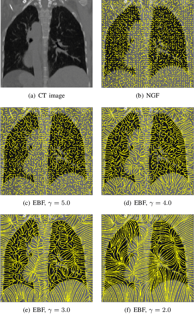
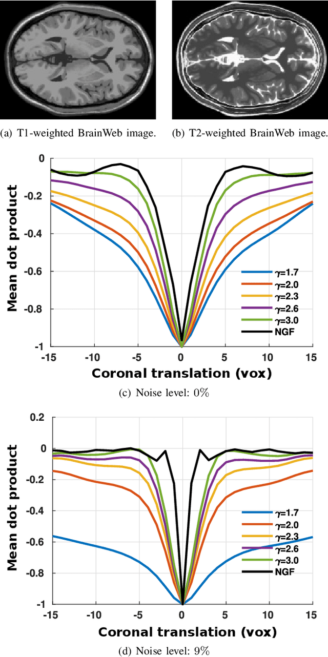
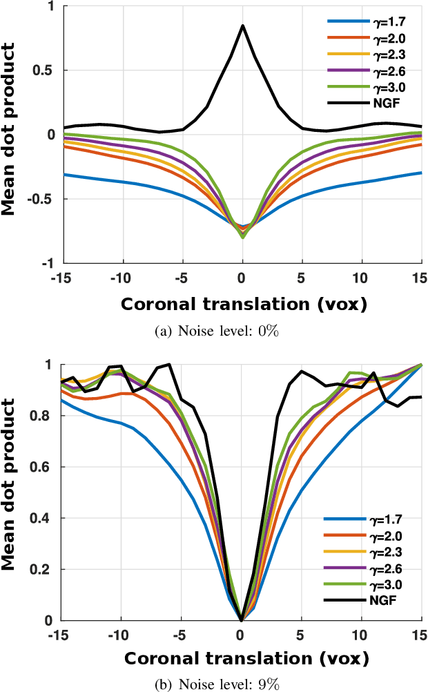
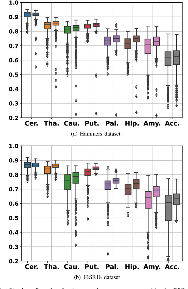
Abstract:In image registration, many efforts have been devoted to the development of alternatives to the popular normalized mutual information criterion. Concurrently to these efforts, an increasing number of works have demonstrated that substantial gains in registration accuracy can also be achieved by aligning structural representations of images rather than images themselves. Following this research path, we propose a new method for mono- and multimodal image registration based on the alignment of regularized vector fields derived from structural information such as gradient vector flow fields, a technique we call \textit{vector field similarity}. Our approach can be combined in a straightforward fashion with any existing registration framework by substituting vector field similarity to intensity-based registration. In our experiments, we show that the proposed approach compares favourably with conventional image alignment on several public image datasets using a diversity of imaging modalities and anatomical locations.
Bone Surface Reconstruction and Clinical Features Estimation from Sparse Landmarks and Statistical Shape Models: A feasibility study on the femur
Jul 07, 2021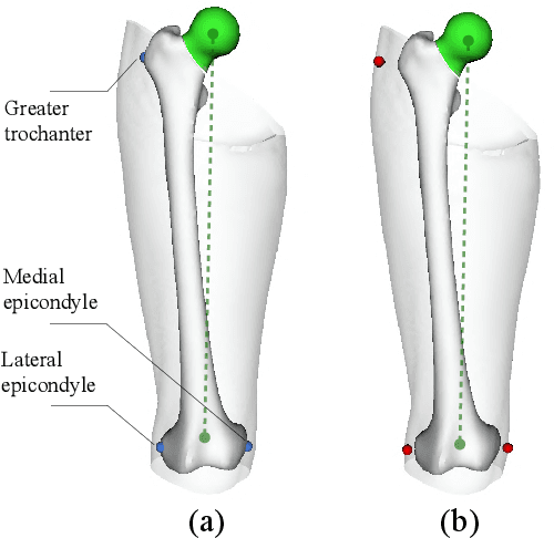


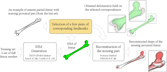
Abstract:In this study, we investigated a method allowing the determination of the femur bone surface as well as its mechanical axis from some easy-to-identify bony landmarks. The reconstruction of the whole femur is therefore performed from these landmarks using a Statistical Shape Model (SSM). The aim of this research is therefore to assess the impact of the number, the position, and the accuracy of the landmarks for the reconstruction of the femur and the determination of its related mechanical axis, an important clinical parameter to consider for the lower limb analysis. Two statistical femur models were created from our in-house dataset and a publicly available dataset. Both were evaluated in terms of average point-to-point surface distance error and through the mechanical axis of the femur. Furthermore, the clinical impact of using landmarks on the skin in replacement of bony landmarks is investigated. The predicted proximal femurs from bony landmarks were more accurate compared to on-skin landmarks while both had less than 3.5 degrees mechanical axis angle deviation error. The results regarding the non-invasive determination of the mechanical axis are very encouraging and could open very interesting clinical perspectives for the analysis of the lower limb either for orthopedics or functional rehabilitation.
 Add to Chrome
 Add to Firefox
 Add to Edge