Bone Surface Reconstruction and Clinical Features Estimation from Sparse Landmarks and Statistical Shape Models: A feasibility study on the femur
Paper and Code
Jul 07, 2021


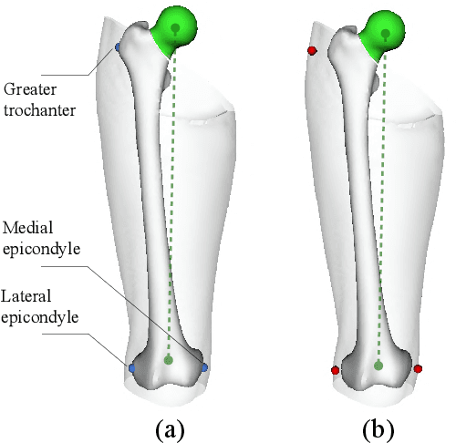
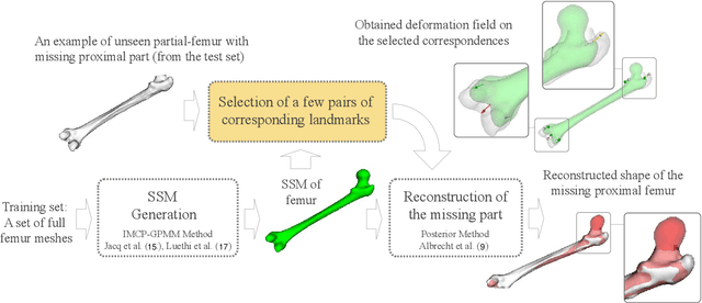
In this study, we investigated a method allowing the determination of the femur bone surface as well as its mechanical axis from some easy-to-identify bony landmarks. The reconstruction of the whole femur is therefore performed from these landmarks using a Statistical Shape Model (SSM). The aim of this research is therefore to assess the impact of the number, the position, and the accuracy of the landmarks for the reconstruction of the femur and the determination of its related mechanical axis, an important clinical parameter to consider for the lower limb analysis. Two statistical femur models were created from our in-house dataset and a publicly available dataset. Both were evaluated in terms of average point-to-point surface distance error and through the mechanical axis of the femur. Furthermore, the clinical impact of using landmarks on the skin in replacement of bony landmarks is investigated. The predicted proximal femurs from bony landmarks were more accurate compared to on-skin landmarks while both had less than 3.5 degrees mechanical axis angle deviation error. The results regarding the non-invasive determination of the mechanical axis are very encouraging and could open very interesting clinical perspectives for the analysis of the lower limb either for orthopedics or functional rehabilitation.
 Add to Chrome
 Add to Firefox
 Add to Edge